Look deep into my eyes…
You’ve probably heard the saying that the eyes are the windows to the soul. While that may a matter of interpretation, what’s definitely true is that an eye doctor can tell you a lot about your health by looking deeply into your eyes.
While most people know the importance of getting regular eye exams in order to screen for vision-threatening diseases like macular degeneration and glaucoma, there are a lot of other conditions that can leave tell-tale signs in your eyes which an ophthalmologist can detect and that can serve as an early warning of potential health problems down the road.
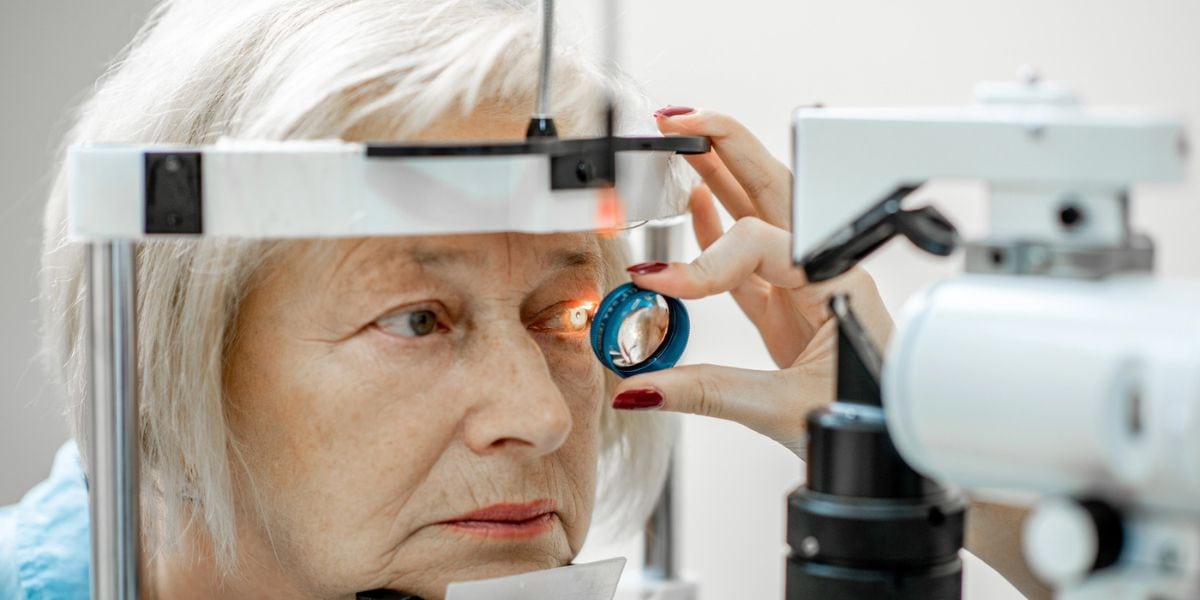
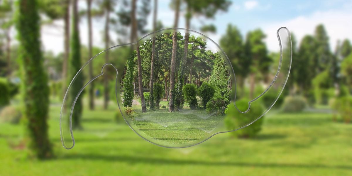
That’s because the blood vessels and nerve cells that reside in the area in the back of your eye, known as the retina, actually reflect what’s going on elsewhere in your body. Early signs of heart disease, diabetes, and even cancerous tumors are often detected through a fundus exam (a dilated eye examination to see the back of your eye).
Your eyes can provide clues linked to Alzheimer’s
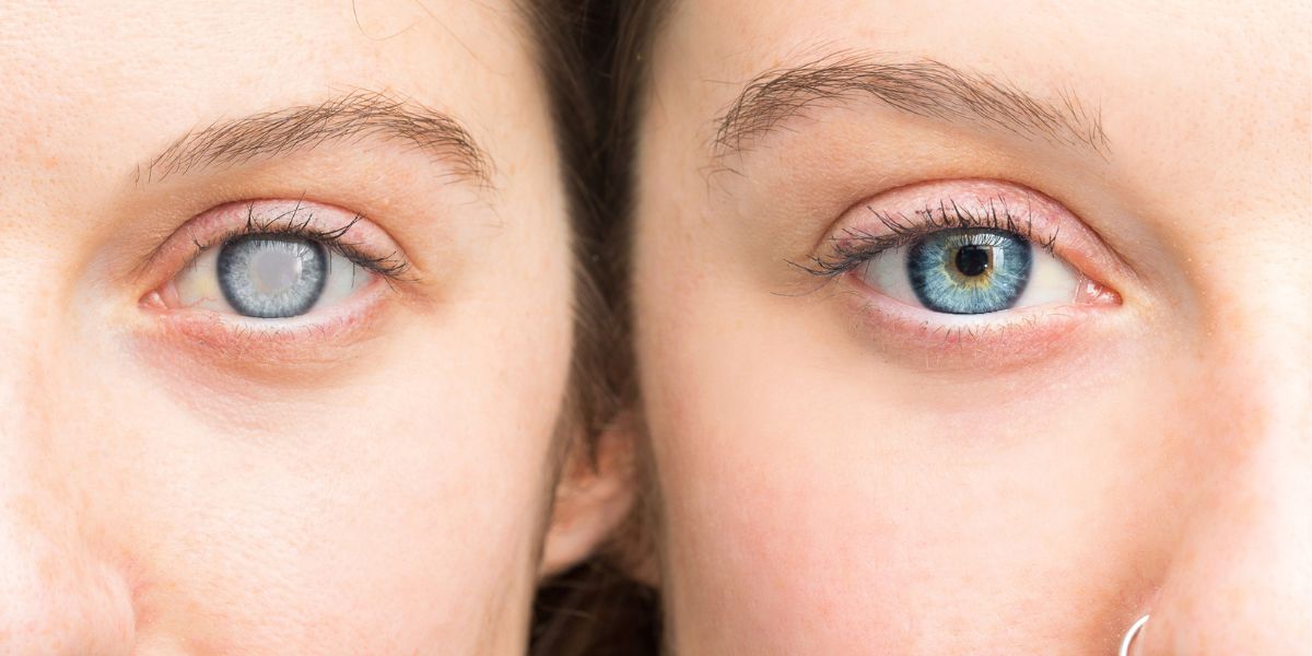
A recent British study shows that your eyes might provide clues to your brain health: specifically, certain changes in your retina have been associated with Alzheimer’s disease.
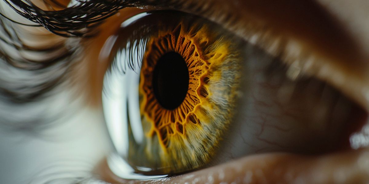
When UK researchers performed retinal scans on the eyes of 120 elderly subjects, they noticed that 25 percent of those with Alzheimer’s had yellow deposits of fat and calcium in the back of the eye known as “hard drusen” compared to just 4 percent of subjects without Alzheimer’s who had hard drusen.
While hard drusen have long been considered pretty harmless, because they rarely impair your vision, they are a sign of oxidative stress. Oxidative stress is basically when there is an imbalance between the number of free radicals and antioxidant defenses in the body. And it is a key factor linked to the development of Alzheimer’s.
Oxidative stress has been implicated in diabetes, hardening of the arteries (aka atherosclerosis), high blood pressure, inflammatory conditions, heart disease, cancer, and neurodegenerative diseases such as Parkinson’s and Alzheimer’s.
Another key finding of the British study was that Alzheimer’s patients had thicker blood vessels in the back of their eyes than the controls. As you might imagine, when your blood vessel walls are thick it’s harder for blood to flow through them, and this means that the eyes (and, by extension, the brain) are receiving less blood flow. And poor circulation is another risk factor causing Alzheimer’s and memory loss.
Can Alzheimer’s be diagnosed with an eye test?
It would be inaccurate to say that a comprehensive eye exam can serve as an Alzheimer’s eye test. It takes a lot more than an eye exam to diagnose Alzheimer’s disease because there are many factors involved in the detection of Alzheimer’s that are outside the purview of eye doctors’ offices. In fact, there is no single Alzheimer test.
Nonetheless, the British study does seem to suggest that early signs of Alzheimer’s are in the eye because it demonstrated that hard drusen on your retina are important biomarkers for increased risk of developing neurodegenerative disorders such as Alzheimer’s and Parkinson’s disease.
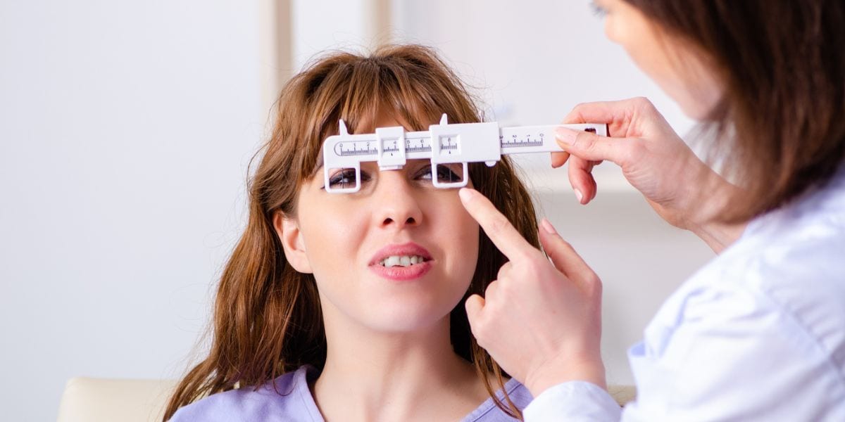
The key takeaway from this study is that having annual eye exams is vitally important for not only screening for eye diseases but also to gain some insight as to your general health and pick up clues to diseases while they’re in their early stages. And, luckily, the same healthy habits that protect you from eye disease can help shield you from dementia.
How to avoid becoming a statistic
At the risk of sounding like a broken record (boy, that sure dates me!) it’s all about adopting healthy dietary choices. Including lots of leafy greens like spinach, kale and collards can not only reduce your oxidative stress but also decrease your risk of glaucoma and dementia because they are rich in nitrate, a compound that your body converts to nitric oxide (NO) which plays a role in increasing blood flow.
And eliminating unrefined “white” carbs like sugar and flour can lower your resting blood sugar and insulin resistance, both of which increase your risk of developing macular degeneration, diabetic retinopathy, and Alzheimer’s disease.
The reason that this ophthalmologist and all of your health care providers give you the same advice is that there’s a ton of research to back up the fact that modifying your diet and lifestyle can lower not just your Alzheimer’s risk but also offset many other disease processes at their earliest stages, leading to a long and healthy life!
Why choose Assil Gaur Eye Institute for your eye care?
The eye care professional team of ophthalmologists and optometrists at Assil Gaur Eye Institute (AGEI) offer world class eye care treating dry eye conditions, cataracts, glaucoma, LASIK and laser eye correction as well as a wide variety of cornea and retinal conditions.
At AGEI, you will experience a state-of-the-art health care facility that brings together revolutionary technologies with experienced vision care professionals. Our goal is to help you achieve your personal best vision.
Please call 866-945-2745 or visit us here to make an appointment online. If you are experiencing any concerning symptoms, contact us immediately to determine the best time to schedule an exam.
At Assil Gaur Eye Institute we take our patients’ safety seriously. Our facility’s Covid-19 patient safety procedures exceed all CDC recommendations. Masks are required in our institutes at all times.
We are conveniently located for patients throughout Southern California and the Los Angeles area at locations in or near Beverly Hills, Santa Monica, West Los Angeles, West Hollywood, Culver City, Hollywood, Venice, Marina del Rey, Malibu, Manhattan Beach, and Downtown Los Angeles, to name a few.