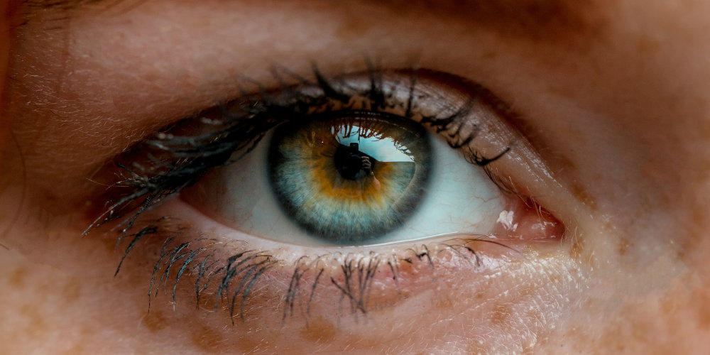
A healthy, intact retina is critical for normal vision. The retina is a thin layer of light-sensitive tissue lining the back of the eye cavity like wallpaper.
Light rays enter the eye through the opening of the iris and converge on the retinal surface where they are converted into electrical impulses. These impulses are carried by the optic nerve to the brain where they are interpreted as images.
When these impulses are interrupted by a retinal condition, the results can be catastrophically disruptive to vision.
There are a number of common retinal diseases that prompt people to see their eye doctor. These include:
Floaters are small spots or what look like strands of hair that appear spontaneously and seem to be floating in your field of vision. They're most noticeable when you stare at a light wall, ceiling, or piece of paper, and they seem to float away if you try to focus on them. Floaters are usually, but not always, harmless signs of aging eyes. In fact, 70% of folks experience floaters at some time.
As we age, the thick gel-like liquid within our eyeballs thins out and can form clumps and strands. These cast a shadow on our retina that is interpreted by our brain as floating specks or lines.
Learn about Floaters
Flashes occur when the gel-like fluid inside our eyes begins to pull away from the retina lining and, in doing so, causes the brain to perceive small bursts or flashes of light. This can be alarming, particularly when it occurs to someone over 45 years old.
Although annoying, floaters and flashes are not usually vision-threatening and do not require treatment. Often floaters decrease and become less bothersome over time. In cases where new floaters and flashes do indicate a more serious condition, such as a retinal tear or retinal detachment, laser treatment or a surgical procedure called a vitrectomy can be used to prevent vision loss.
Learn more about Flashes
The macula is a small area in the central retina with special cells that allow you to see fine details so you can make out faces, read or drive, for example. A macular hole is typically seen in people over age 55 and, more often, in women. This is a condition that requires vitrectomy surgery for treatment.
Learn more about Macular Holes
A very common and relatively harmless condition called macular pucker or epiretinal membrane is a thin translucent layer of connective tissue that slowly develops over the macula. Over time, this sheet can contract and wrinkle or pucker, leading to blurred central vision and straight lines appearing wavy.
There are no eye drops, medications, or supplements to treat epiretinal membranes. Typically they are observed over time, as they grow very slowly. If they begin to cause visual symptoms, vitrectomy surgery is performed to remove them.
Learn more about Macular Puckers
There are a number of serious retinal conditions that require timely medical intervention in order to prevent permanent loss of vision. These include:
AMD is the age-related deterioration of the center of the retina, known as the macula. The macula is responsible for fine focus that lets you distinguish faces, read, and drive. AMD is one of the leading causes of decreased vision in Americans over age 50.
Symptoms of AMD include blurred central vision with dark spots, colors appearing dim, distorted central vision causing straight lines to appear curved, and making words difficult to read. AMD typically spares peripheral vision.
There are two forms of AMD:
Dry macular degeneration is the most common form of AMD. It progresses slowly, is mostly associated with mild visual changes, and is treated with a dietary supplement and timed observation.
Wet macular degeneration is the more severe, less common stage of AMD, can progress rapidly, causing significant, permanent central vision loss, and requires timely treatment with intraocular injectable medications.
Learn more about Macular Degeneration
A retinal detachment is a vision-threatening emergency in which the retina pulls away from the inside eyewall. It's an emergency because the retina gets oxygen and nourishment from the blood vessels located in the underlying tissue on the eyewall. The longer a retinal detachment goes untreated, the greater your risk of permanent vision loss.
A retinal detachment often starts with a small tear or hole in the retina, called a retinal tear. This is concerning because it can expand and allow the jelly-like fluid within your eye to seep through and build up behind the retina. Over time, this fluid build-up can cause the retina to peel off from the eyewall-like wallpaper.
Symptoms of a retinal detachment include the sudden appearance of flashes of light, floaters, and a curtain-like shadow blocking your visual field. A retinal detachment is an emergency. In order to preserve as much vision as possible, your eye surgeon needs to reattach the delicate retina tissue to the wall quickly.
Learn more about Retinal Tears and Retinal Detachment
Long-term elevated blood glucose levels damage small blood vessels responsible for nourishing the retina. This causes the vessel walls to weaken and bulge, making them leak fluid underneath the retinal surface.
As tiny blood vessels get damaged and begin to close off, the retina secretes special growth factors that cause abnormal new blood vessels to sprout on the retinal surface and within the gel-like fluid within your eye. This process is called proliferative diabetic retinopathy.
Unfortunately, these new vessels also have weak walls that can leak and bleed into your eye, causing blurred vision, scarring, or dangerously high eye pressure. They form scar tissue that can pull away from the eyewall, causing retinal detachment and permanent vision loss.
Learn more about Proliferative Diabetic Retinopathy
In advanced diabetes, elevated blood glucose levels damage the blood vessels nourishing the retina. This causes them to leak fluid underneath the retinal surface. When the leaking involves the central retinal area, known as the macula, this is known as macular edema.
Macular edema can eventually destroy the macula, resulting in partial or total vision loss. In fact, macular edema is the most common cause of vision loss among people with diabetic retinopathy.
The Central Retinal Artery is the key artery responsible for carrying oxygen-rich blood to the nerve cells that make up the retina lining the back of your eye. If this artery gets blocked by a blood clot or a bit of cholesterol, it robs the retinal cells of oxygen and they begin to die off. The blockage can last for seconds, minutes, or can even be permanent, in which case it will lead to permanent vision loss.
The typical symptom of a CRAO is the sudden onset of severe vision loss. A CRAO requires treatment within 4 to 6 hours because oxygen must be quickly restored, or nerve cells will die off.
Treatment options include hyperventilation with highly concentrated oxygen, medications, or surgery to decrease eye pressure, in an attempt to dislodge a blood clot or cholesterol plug in the artery.
Learn more about Central Retinal Artery Occlusion
Sometimes the main vein responsible for draining deoxygenated blood from the retina becomes partially or completely blocked due to a clot or decreased blood flow. This can cause pressure build-up in the vessels, making them leak blood (known as retinal hemorrhages) and fluid underneath the central retina (macular edema).
CRVO symptoms include blurred vision that waxes and wanes. Severe CRVO can cause pain, redness, and increased ocular pressure. The body creates new abnormal blood vessels as it tries to create new drainage pathways for de-oxygenated blood.
Treatment options for CRVO include administering special medications (called anti-VEGF medicines) directly into the eye via injection or performing laser treatments to cauterize vein leaks. More common than CRVO are occlusions of smaller feeder branch retinal veins (BRVO). People over 50, smokers, diabetics, and folks with high blood pressure are at greater risk of developing BRVO.
Learn more about Central Retinal Vein Occlusion (CRVO)
The AGEI staff includes a highly skilled retina specialist Dr. Svetlana Pilyugina or “Dr. P”, as she is known to her patients. Dr. P is a Stanford-educated and fellowship-trained ophthalmologist. She is board certified in diseases and surgery of the vitreous and retina.
Dr. Pilyugina has extensive experience treating floaters, flashes, and all retinal conditions. To schedule an appointment, call 866-945-2745 or click here to make an appointment online.
As a member of an elite group of only 3000 retina-vitreous specialists in the United States, Stanford trained Dr. Pilyugina brings to AGEI a unique skill set in the treatment and surgery of retinal disease. Her academic credentials include numerous research papers, conference presentations, medical publications, and clinical trials.