Cataract Lens Replacement Failure: What Can You Do About It?
Cataract surgery is one of the most common surgeries worldwide. In the United States alone, more than 3.5 million cataract surgeries are performed annually. Globally, the number reaches over 28 million procedures a year. About 90% of people develop cataracts by age 65. Like other age-related degenerative conditions, cataract risk increases with age.
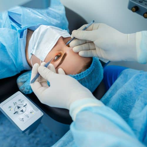
Cataract surgery restores clear vision and becomes necessary when the eye’s natural lens becomes cloudy and impairs vision. The surgery removes the cloudy lens (called cataract extraction) and replaces it with a clear artificial lens called an intraocular lens.

Outpatient cataract surgery takes about 20 minutes, with minimal recovery time and generally no side effects or serious complications. It has a success rate of over 95%. Aftercare is usually just a follow-up appointment and antibiotic eye drops for several days.
With premium cataract surgery, that clear lens can provide amazing vision correction and even eliminate the patient’s need for reading glasses or contact lenses.
Residual refractive error and ocular surface disease
Two of the most common reasons patients feel disappointed after cataract surgery have nothing to do with the lens itself failing – they come down to residual refractive error and ocular surface disease, and both are worth understanding before you assume something went seriously wrong.
Residual refractive error simply means the eye did not land exactly where the surgical plan intended. Even with world-class preoperative measurements, the eye is a biological system, and it heals on its own terms. A small amount of remaining nearsightedness, farsightedness, or astigmatism can leave vision feeling soft or slightly off. The good news is that this is one of the most correctable outcomes in all of eye care. Depending on the degree of residual error and your corneal health, options can include laser enhancement, a lens exchange, or a simple glasses prescription for specific tasks. The key is catching it early with precise diagnostic tools, not waiting and wondering.
Ocular surface disease, most often dry eye, is the other major culprit. Cataract surgery temporarily disrupts the corneal nerves responsible for triggering tear production, and for some patients that disruption lingers for weeks or even months. When the tear film is unstable, light scatters unevenly across the eye’s surface and vision fluctuates throughout the day. Many patients describe it as blurry vision that comes and goes, which can be frustrating when you expected crisp, clear sight right away.
Aggressively treating the ocular surface before and after surgery dramatically reduces this risk. At Assil Gaur Eye Institute, dry eye evaluation is built into the premium cataract surgery process because an unhealthy tear film can also skew the measurements used to select your lens, compounding the problem before the procedure even begins.
Why do intraocular lens implants stop working?
Intraocular lenses used in cataract and RLE (refractive lens exchange) surgery are designed to last a lifetime. However, about 5% of patients report dissatisfaction, and about 3% undergo a second procedure called an IOL exchange, or intraocular lens exchange.

Several reasons and risk factors can explain why an IOL can go bad. However, the more experienced the surgeon, the fewer complications their patients tend to have during cataract surgery. You want your surgeon prepared to handle potential issues, such as performing a vitrectomy if your eye’s capsular bag integrity is compromised. If this is not possible, they need to know how to place the new IOL in the sulcus or anterior chamber.
The causes for needing IOL exchange surgery include:
The patient is not a good candidate
Sometimes, pre-existing problems such as significant macular degeneration (part of the retina), prior ocular surgery (such as an Nd:YAG posterior laser capsulotomy), and lazy eyes can preclude a patient from getting many IOLs.
A surgeon with deep knowledge of problematic eye conditions and the characteristics of all IOLs on the market can help patients avoid potential problems.
Poor quality intraocular lenses
Some patients who come to AGEI for this type of surgery have artificial lenses implanted with poor optics (blurry vision and halos), leading to poor visual outcomes. This is usually because the polymer used to make the IOL does not match the individual’s cornea.
The patient often experiences “Vaseline Vision” (dysphasia). A new lens typically brings about excellent vision.
Poor lens choice for the patient’s cornea
Not every cornea is the same, and selecting the wrong intraocular lens power for a patient’s unique corneal shape is one of the more preventable causes of dissatisfaction after cataract surgery. When the lens power doesn’t match the cornea’s steepness or curvature, patients wake up after surgery still needing significant glasses correction, which defeats the purpose of choosing a premium IOL in the first place.
Corneal irregularity is a particularly common culprit. Eyes with steep or asymmetric corneas, conditions like keratoconus, or significant astigmatism require precise biometry measurements and carefully chosen power-calculation formulas. Standard formulas designed for average corneas can produce meaningful errors when applied to irregular ones, leaving patients with blurry or distorted vision that no amount of glasses adjustment fully corrects.
Post-LASIK and post-refractive surgery eyes present an even greater challenge. Prior laser treatment permanently alters the corneal curvature and changes how light refracts through the eye. Standard IOL calculation formulas were not built with these altered corneas in mind, which is why patients who had LASIK years ago and now need cataract surgery face a higher risk of lens power miscalculation if their surgeon does not account for their refractive history. Clinical research published by the NCBI confirms that wrong lens implant errors in post-refractive eyes remain one of the most studied and difficult-to-prevent complications in cataract surgery.
At AGEI, advanced intraoperative measurement technology allows our surgeons to verify lens selection in real time during surgery itself, before the incision is closed. If measurements suggest the chosen lens will not deliver the precision outcome the patient expects, the lens can be swapped on the spot. This level of surgical precision is what separates a world-class cataract program from a high-volume, one-size-fits-all approach.
For patients who have already experienced a corneal mismatch after surgery elsewhere, the solution can sometimes be as straightforward as our LASIK surgery for farsightedness, which fine-tunes residual refractive error without replacing the lens entirely.
Posterior capsular opacification (PCO)
The most common issue associated with IOLs is posterior capsule opacification, also known as a “secondary cataract.” This occurs when a part of the lens capsule thickens, causing blurred vision. It can occur months to years after surgery.
This isn’t technically lens failure, and the new IOL hasn’t become cloudy. PCO occurs when epithelial cells from the natural lens, left behind during surgery, proliferate and migrate onto the posterior capsule (the thin membrane that holds the intraocular lens in place).
This gradually clouds the capsule behind the IOL, which can cause blurred vision, glare, or halos, similar to the original cataract symptoms. Studies suggest PCO develops in roughly 20 to 40 percent of patients within two to five years of cataract surgery, making it by far the most frequently encountered long-term complication.
Fortunately, this is easily treated with a straightforward laser procedure. The standard treatment is an in-office procedure called Nd:YAG laser capsulotomy, in which a precisely focused laser beam creates a small, clear opening in the clouded capsule to restore the light path to the retina. The procedure typically takes only a few minutes, requires no incisions, and carries no downtime. Most patients notice a meaningful improvement in clarity within 24 hours, and the success rate is consistently reported above 95 percent. Once the capsulotomy is performed, PCO does not recur in that same area, so a single treatment is almost always all that is needed.
Lens dislocation
A dislocated intraocular lens, which generally occurs more than three months after cataract surgery, can cause blurry vision, halos around bright lights, or double vision.
Some of the causes of this postoperative complication include:
Inherited connective tissue disorders (e.g., Marfan syndrome, hyperlysinemia, homocystinuria, Ehlers-Danlos syndrome), capsular contraction
Surgical trauma
Repeated eye infections
High myopia (severe nearsightedness)
Facial or eye injury
Eye inflammation
Conditions that make the capsular bag unstable, such as pseudoexfoliation syndrome
Family history of Marfan’s syndrome, a genetic disorder that affects the connective tissue in your eyes and other parts of the body
A history of retina surgery or injection therapy
If a lens dislocates far into the back of the eye, a retinal surgeon must perform a vitrectomy to ensure the retina is not damaged before removing the lens.
Intolerable glare and light sensitivity
Glare and light sensitivity after cataract surgery usually fall under a clinical category called dysphotopsia, which comes in two distinct forms worth understanding before surgery.
Positive dysphotopsia refers to unwanted light phenomena that patients actually see: glare, starbursts radiating from headlights, halos around streetlamps, and streaks of light that make night driving genuinely difficult. Negative dysphotopsia is the opposite experience, a dark arc or crescent-shaped shadow that appears in the peripheral vision, typically in the temporal field. Both can be disorienting, and both are more common when the lens implanted does not suit the patient’s specific eye anatomy.
Those who experience painful glare and light sensitivity generally receive a lens mismatched to their eye shape. Multifocal IOLs, for example, split incoming light across multiple focal points, which is excellent for reading independence but can intensify halos and starbursts in patients with naturally large pupils or corneas with pre-existing irregularities. A thorough pre-operative evaluation should flag these risk factors before choosing any lens.
A mechanical cause is also easy to overlook. When the haptics, the small arms that anchor the lens inside the capsular bag, exert uneven pressure on the bag wall, they can create subtle folds called capsular striae. Those striae distort the path of incoming light and produce starburst patterns that patients often mistake for a defective lens. In reality, the lens itself may be fine; the problem is how it is sitting inside the eye.
This can typically be prevented by seeing a highly experienced surgeon who matches lens design to corneal shape, pupil size, and lifestyle needs before the procedure begins.
Untreated inflammation
Some degree of inflammation after cataract surgery is completely normal. Your immune system recognizes the procedure as a disruption and responds accordingly, which is why surgeons routinely prescribe anti-inflammatory eye drops as part of your post-operative care plan.
The problem arises when that inflammatory response goes unchecked. Cystoid macular edema (CME) is one of the more serious consequences of untreated post-surgical inflammation. It occurs when fluid accumulates in the macula, the central region of the retina responsible for sharp, detailed vision. Patients typically describe their vision as blurry, washed out, or distorted, sometimes weeks after surgery seemed to go smoothly. CME is more likely in patients who have diabetes, a history of uveitis, or who stopped using their prescribed drops too soon.
Beyond CME, persistent inflammation can cause corneal swelling, elevated eye pressure, and, in prolonged cases, damage to surrounding ocular structures. Watch for redness that doesn’t settle, light sensitivity, and vision that worsens rather than improves in the days after surgery.
The good news is that when caught early, post-surgical inflammation responds well to treatment. Prescription steroid and non-steroidal anti-inflammatory drops are the first line of defense, and most cases resolve fully with consistent use. More stubborn cases may require a corticosteroid injection near the eye. The critical factor is compliance: finishing the full course of prescribed drops, even when the eye feels fine, is one of the simplest ways to prevent a manageable response from becoming a serious complication.
Endophthalmitis or infection
Though not directly a cataract lens problem, an infection after cataract surgery can negatively affect the overall outcome. If the infection becomes severe, the IOL may need to be removed.
Retinal detachment after cataract surgery

Retinal detachment is one of the more serious complications that can follow cataract surgery, and it is worth understanding even though it is relatively uncommon. The retina is the thin layer of light-sensitive tissue lining the back of your eye. During cataract surgery, the natural lens is removed, and that process can sometimes disturb the gel-like vitreous fluid inside the eye. When the vitreous pulls away from the retina, it can create a small tear. If fluid seeps through that tear and lifts the retina away from the underlying tissue, a detachment occurs.
The risk is higher for people who are highly nearsighted, have a history of retinal problems, or have experienced any surgical complications during their cataract procedure. You can read more about related retinal conditions and how they develop over time. Watch for a sudden increase in floaters, flashes of light, or a shadow creeping across your peripheral vision. These are not normal parts of recovery and should be treated as a same-day emergency.
The good news is that retinal detachment is treatable, especially when caught early. Surgical repair, whether through laser treatment, cryotherapy, or a more advanced procedure like vitrectomy, can reattach the retina and preserve vision. At Assil Gaur Eye Institute, retinal specialist Dr. Svetlana Pilyugina brings world-class precision to the surgical management of retinal tears and detachments, giving patients the best possible chance at full visual recovery. Prompt action is everything with this complication, so never wait to report sudden changes in your vision after cataract surgery.
Elevated eye pressure and other post-surgical complications
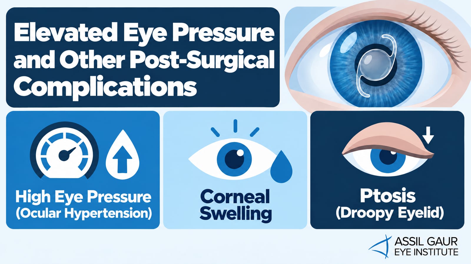
Beyond the complications covered above, a handful of other post-surgical issues can affect how well your IOL performs and how comfortable your vision feels day to day. Understanding what these are makes it much easier to catch them early and get the right care.
High eye pressure (ocular hypertension): After cataract surgery, fluid drainage inside the eye can temporarily become sluggish, causing intraocular pressure to spike. If that pressure stays elevated without treatment, it can damage the optic nerve and lead to glaucoma. Your surgeon will typically monitor your pressure closely in the days following surgery and may prescribe eye drops to bring it back to a safe range. Patients who already have a history of glaucoma are at higher risk and need especially careful follow-up.
Corneal swelling: The cornea can swell after surgery as a reaction to the procedure itself. This usually resolves on its own within a few days, but in some cases it lingers and noticeably blurs vision. Anti-inflammatory drops are the standard first response, and most patients recover fully with consistent use.
Ptosis (droopy eyelid): A small number of patients notice that one eyelid sits lower than before surgery. This happens because the speculum used to hold the eye open during the procedure can temporarily stretch the muscle that lifts the lid. It often resolves within weeks, though persistent cases may benefit from a targeted oculoplastic evaluation.
Catching any of these complications early is key. If something feels off after your procedure, prompt follow-up with a world-class surgical team gives you the best chance at an enviable outcome. Explore more cataract surgery insights on the AGEI blog to stay informed throughout your recovery.
Call now or book your appointment online.
Sophisticated instruments allow AGEI to gather accurate measurements and prevent artificial lens implant failures
It would be an understatement to say that accurate preoperative and operative measurements for artificial lens implants are essential to an excellent visual outcome.
Assil Gaur Eye Institute has invested in cutting-edge technology to achieve the most accurate eye and lens measurements possible. These include:
Intraoperative Aberrometry (ORA)
ORA is a tool that allows surgeons to take refractive measurements in the operating room during surgery, both aphakic (without a lens) and pseudophakic (with a lens in the eye).
Our own Dr. Kerry Assil helped Alcon develop and test this technology and was the first to use it worldwide. The ORA system allows our surgeons to confirm that they have selected the ideal IOL for optimal vision correction and the perfect lens placement location in the eye. Because a large percentage of IOL exchanges are due to incorrect IOL power, the ORA system is essential.
Typically, surgeons have no choice but to use the best lens based on pre-operative measurements. However, because AGEI uses ORA technology and has a vast inventory of almost every power for each IOL implant, they can make any needed correction while the patient is still in the operating room.
IOL Master
IOL Master is the gold standard for optical biometry (a highly accurate, non-invasive, automated method for measuring the eye’s anatomical characteristics). It helps with intraocular lens power calculations by measuring the eye’s anatomical features, such as axial length, corneal curvature, and anterior chamber depth. All these are essential for calculating the correct power of intraocular lenses (IOLs) during cataract surgery.
LensAR Femtosecond Laser
The LensAR Femtosecond Laser is an advanced laser system used in ophthalmology, specifically for cataract surgery. Also known as laser-assisted cataract surgery, this laser performs critical steps in the laser eye surgery process with high precision, enhancing the safety and effectiveness of the procedure.
This system captures ten images from different angles, giving our surgeon a 3D image of the cataract and revealing its thickness and density.
The femtosecond laser softens the cataract by making highly accurate, reproducible cuts. This leads to much better outcomes than without it. It also reduces surgical time by automating and optimizing key steps. The laser’s precision maximizes safety by minimizing the risk of complications, often leading to quicker recovery.
Why trust AGEI to correct any issues you may have from cataract surgery or refractive lens exchange surgery
With over 30 years of experience, Dr. Kerry Assil and his partner, Dr. Avneet K. Sodhi Gaur, are among the most experienced cataract surgeons in the United States. Dr. Assil has performed over 70,000 eye surgeries and authored over 100 textbook chapters on vision problems, as well as articles on refractive errors and cataract surgery.
If your IOL implant is no longer helping your vision, don’t despair. Often, patients seeking a revision turn to AGEI because they want their IOL to provide clear vision.
Assil Gaur Eye Institute has assembled a team of top ophthalmologists from across the country who provide patients with the highest-quality specialist eye care in the United States.
In keeping with AGEI’s founding principles, eye surgeons keep their clinics comfortable and familiar, much like family-run medical practices once were.
We offer state-of-the-art eye exams, premium cataract surgery, and refractive lens exchange (RLE) using the best-chosen lens for each patient. These lenses include extended depth of focus IOLs, multifocal IOLs, and monovision / monofocal lenses (often to correct for distance vision or near vision).
In addition to refractive lens exchange and cataract surgery, the ophthalmology experts at AGEI are nationally recognized for their eye care and treatment options for a full range of conditions, including eye conditions such as hyperopia, glaucoma, retinal detachment (and other vision loss retinal procedures), cornea conditions, floaters and flashes, dry eye, presbyopia, astigmatism (using a toric lens), farsightedness, myopia, nearsightedness, and other vision correction procedures (refractive surgery) such as LASIK surgery, PRK, and blurred vision.
There is a solution, and we invite you to contact us for a consultation. Please call (866) 945-2745 or make an appointment online.
Our eye centers are conveniently located throughout Southern California and the Los Angeles area, including Beverly Hills, Santa Monica, West Los Angeles, West Hollywood, Culver City, Hollywood, Venice, Marina del Rey, Malibu, Manhattan Beach, and Downtown Los Angeles.
Call now or book your appointment online.
Intraocular Lens (IOL) FAQs
Can a lens fail after cataract surgery?
Though rare, a lens can fail or have complications after cataract surgery. The failure can manifest differently depending on the intraocular lens (IOL) issue.
Can a cataract lens be replaced more than once?
Yes, an intraocular lens (IOL) implanted during lens replacement surgery can be replaced more than once if necessary. Although rare, doctors call the IOL replacement procedure an IOL exchange. Doctors typically perform it when the original lens causes significant visual problems or complications that cannot be corrected otherwise.
What should I do if I experience blurry vision after cataract lens implantation?
If you experience blurry vision after cataract lens implantation, it’s essential to determine the underlying cause. Blurry vision can occur immediately after surgery as the eye heals, but if it persists, don’t ignore it. See your ophthalmologist.
How can I tell if my intraocular lens (IOL) has dislocated or displaced? What are the signs of lens dislocation after cataract surgery?
If your intraocular lens (IOL) becomes dislocated or displaced after cataract surgery, several signs and symptoms can indicate this problem. IOL dislocation can occur shortly after surgery or even years later. Here are some visual disturbances to look for:
- Blurry or decreased vision
- Double vision (Diplopia)
- Visual distortions such as glare or halos around lights, especially in low-light conditions like driving at night; ghosting or shadowing around objects; or changes in the shape or size of images, sometimes described as a “wavy” or “warped” appearance.
- Feeling of “lens movement” in the eye
- A sudden change in your prescription
- Visible IOL edge
- Astigmatism
- Persistent eye discomfort
-
Cataract Surgery Complications
https://eyewiki.aao.org/Cataract_Surgery_Complications -
Vitreous Hemorrhage
https://eyewiki.aao.org/Vitreous_Hemorrhage -
Visual Acuity Outcomes and Complications after Intraocular Lens Exchange
https://doi.org/10.1016/j.ophtha.2023.10.021 -
Ten Cataract Surgery Side Effects, and How to Cope – American Academy of Ophthalmology
https://www.aao.org/eye-health/tips-prevention/side-effects-cataract-surgery-complications-cope

Written by The AGEI Educational Team

Medically Reviewed by Dr. Kerry Assil