Keratoconusin Los Angeles
in Los Angeles
What is keratoconus?
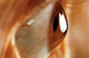
Keratoconus is an eye condition in which the cornea (the clear outer layer of the eye) progressively thins and becomes irregularly (coned) shaped.
The abnormal curvature of the cornea causes the cornea to bulge and prevents the light entering the eye from focusing correctly on the retina, and causes vision to become increasingly distorted. As the condition worsens and the front of the cornea expands, you begin to experience myopia(nearsightedness) and develop irregular astigmatism.
What is keratoconus?
Keratoconus is a progressive eye condition in which the cornea, the clear dome-shaped surface at the front of your eye, gradually thins and begins to bulge outward into a cone-like shape. Normally, the cornea has a smooth, rounded curve that helps focus light precisely onto the retina. When keratoconus develops, that curve becomes irregular, scattering light as it enters the eye and producing blurry, distorted vision that glasses and standard contact lenses can struggle to correct.
The condition typically appears in the teenage years or early twenties and can progress steadily through a person’s thirties before stabilizing. It affects roughly one in every 2,000 people, though researchers believe the true number may be higher because mild cases are sometimes missed without advanced corneal imaging. Keratoconus usually develops in both eyes, although one eye is often more affected than the other.
Catching keratoconus early matters because it is a progressive condition. If left unmanaged, it can progress from minor visual disturbances to significant vision impairment that interferes with everyday activities like reading, driving, and recognizing faces. The good news is that with world-class care and advanced diagnostic tools, doctors can detect keratoconus at its earliest stages and treat it with precision before it seriously compromises your quality of life. Understanding what keratoconus is and how it behaves is the first step toward protecting your vision for the long term.
What are the symptoms of keratoconus?
Signs and symptoms of keratoconus can include:
Blurred vision or distorted vision with difficulty seeing near and far
Increasing light sensitivity and glare, and halos, which can compromise night driving
Unstable vision leading to frequently changing eye prescriptions
Increased dry eye symptoms while wearing conventional contact lenses
Monocular diplopia, meaning double vision that persists even when one eye is closed, caused by the irregular shape of the cornea bending light unevenly across the retina
Ghosting around letters, images, or lights, which is one of the earliest and most frustrating visual complaints patients describe before a formal keratoconus diagnosis is made
Beyond what patients notice day to day, a world-class keratoconus specialist will also look for clinical signs visible only during a slit-lamp examination. Two of the most telling are the Fleischer ring, a brownish iron deposit that forms in a circular pattern at the base of the cone, and Vogt’s striae, fine vertical stress lines that appear deep within the corneal tissue as the cone steepens and stretches. These findings help confirm the diagnosis and give your care team a clearer picture of how far the condition has progressed.
Because keratoconus tends to advance gradually, many patients spend years cycling through updated glasses and contact lens prescriptions before identifying the underlying cause. If you are noticing any of these symptoms, a comprehensive corneal evaluation is the essential first step toward protecting your vision and exploring the advanced treatment options available at Assil Gaur Eye Institute.
What causes keratoconus?
Keratoconus is a progressive condition that is more prevalent in teenagers and adults in their 30s. It can progress gradually or quickly, and usually affects both eyes. Unfortunately, the cause of keratoconus is not fully understood. There is a known genetic connection since ten percent of keratoconus sufferers have a parent with the condition. It’s also often seen in patients who suffer from retinitis pigmentosa, Down syndrome, Ehlers-Danlos syndrome, hay fever, and asthma.
Some medical researchers theorize that an enzyme imbalance in the cornea makes it vulnerable to free-radical damage, weakening the corneal tissue and causing it to bulge. This theory has gained significant clinical support. Research suggests that the corneal stroma in keratoconus eyes contains lower levels of protective antioxidant enzymes, including superoxide dismutase and catalase. Without adequate antioxidant defense, oxidative stress builds up within the corneal tissue, breaking down the collagen fibers that normally keep the cornea firm and dome-shaped. Over time, this structural breakdown allows the cornea to thin and progressively bulge forward into the irregular cone shape that defines the condition.
This biochemical explanation also helps clarify why certain lifestyle and environmental factors appear to accelerate keratoconus progression. Chronic eye rubbing, for example, is strongly associated with worsening of the condition, likely because repeated mechanical trauma generates additional oxidative stress in already vulnerable corneal tissue. Prolonged UV exposure and poorly managed eye allergies, which trigger habitual rubbing, are similarly linked to faster progression. Understanding these triggers is important because avoiding them is one of the few ways patients can actively slow the disease before advanced treatment becomes necessary.
What are the risk factors for Keratoconus?
Frequent vigorous eye rubbing is thought to increase your risk of developing keratoconus. Additionally, overexposure to ultraviolet rays, improper contact lens wear, and chronic eye irritations may increase your odds of developing keratoconus.
Can keratoconus be prevented?
Because genetics play a significant role in keratoconus, there is no guaranteed way to prevent the condition entirely. However, you can take practical steps to reduce your risk or slow its progression, especially if you already know you carry some of the risk factors discussed above.
The single most important habit to break is eye rubbing. Chronic, vigorous rubbing puts repeated mechanical stress on the cornea and is strongly associated with both the onset and worsening of keratoconus. If you find yourself rubbing your eyes frequently, it is worth talking to a specialist about why. Allergies are one of the most common culprits, and managing them properly with antihistamine eye drops or other treatments can make a real difference in protecting your corneal health.
UV exposure is another factor worth taking seriously. Wearing quality sunglasses that block UVA and UVB rays helps shield the cornea from oxidative damage that may contribute to corneal weakening over time. This is especially relevant in a sun-drenched city like Los Angeles, where year-round UV exposure is simply part of daily life.
Perhaps the most powerful preventive tool available is early detection. Routine eye care and advanced corneal mapping can identify subtle changes in corneal shape long before symptoms become noticeable. Catching keratoconus early opens the door to treatments that can stabilize the cornea and preserve your vision for the long term. If you have a family history of keratoconus or notice any changes in your vision, scheduling a comprehensive evaluation sooner rather than later is always the right move.
How is keratoconus diagnosed?
Typically, doctors diagnose keratoconus during an eye exam when they notice an irregularly shaped cornea.
As part of a comprehensive eye exam, our eye doctor measures your cornea’s shape to screen for keratoconus. With annual eye exams, we can detect and track subtle changes in keratoconus in its early stages.
At AGEI, we use state-of-the-art corneal topography, which involves imaging the cornea to analyze its surface shape and characteristics in just a few seconds. We also perform corneal tomography using Scheimpflug imaging, which goes a step further by mapping the full thickness of the cornea from front to back – giving our specialists a far more complete picture of how the cornea is changing over time. This is especially valuable for catching keratoconus in its earliest stages, before symptoms become noticeable.
Pachymetry is another essential part of our diagnostic process. This quick, painless measurement tells us exactly how thin your cornea has become at any given point, which helps our team determine how advanced the condition is and which treatment path makes the most sense for you.
A slit-lamp examination rounds out the evaluation, allowing our doctors to inspect the cornea under high magnification and look for telltale physical signs of keratoconus, such as Fleischer’s ring, Vogt’s striae, or corneal scarring. Together, these advanced diagnostic tools give our world-class specialists the precision they need to build a treatment plan tailored specifically to your eyes.
Call now or book your appointment online.
What are the treatments for early keratoconus?
Keratoconus has several stages, starting with early diagnosis. The earlier you are diagnosed, the greater the chance we can often easily correct visual issues with glasses or special contact lenses.
Early treatment options, which can often provide excellent quality vision and avoid vision loss for many years, include:
Rigid gas-permeable keratoconus lenses
Rigid gas-permeable lenses are made of specialized plastic that lets oxygen through. These lenses reshape your cornea’s curvature to correct your vision while wearing them.
Custom Toric soft contact lenses
While these are usually more comfortable than rigid gas-permeable contact lenses, they are best for individuals with more astigmatism.
Scleral contact lenses
These are oversized contact lenses whose outer rim extends to the white of your eye, known as the sclera. These lenses vault over the cone-shaped part of your cornea without touching its surface, thanks to a special fluid-filled reservoir that lies over the cone-shaped cornea and restores clear vision comfortably.
Finding properly fitting corrective lenses is crucial. Poorly fitting lenses can cause corneal scarring and intolerance to any lens. At AGEI, our experts can help you choose the lens option that fits your needs and provides the best vision and comfort.
Intermediate-stage keratoconus: what are your options?
When glasses and standard contact lenses are no longer giving you the sharp, stable vision you need, but your keratoconus has not yet progressed to the point where surgery is the only path forward, you are in what specialists call the intermediate stage. This is a critical window of opportunity, and the good news is that several advanced options are worth knowing about.
Corneal collagen crosslinking (CXL) is the standout treatment at this stage. It uses a precise combination of riboflavin eye drops and controlled UV light to strengthen the collagen fibers in your cornea, making it more resistant to further bulging and slowing progression.
Alongside CXL, your eye care team may recommend advanced lens options designed specifically for irregular corneas. Hybrid contact lenses pair a rigid center with a soft outer skirt, giving you the crisp optics of a rigid lens with far more wearing comfort. Piggyback lens systems layer a rigid lens over a soft lens for a similar effect. Both approaches can restore genuinely functional vision while your cornea stabilizes after crosslinking.
The key takeaway is that intermediate-stage keratoconus responds well to world-class, precision-driven care. Acting during this window can slow or even halt progression, protecting your vision and reducing the likelihood of needing more invasive treatment later. Explore more eye health resources to stay informed about your options.
Corneal Collagen Crosslinking for Keratoconus
Early detection and a treatment called Collagen Crosslinking (CXL) can help manage the progression of intermediate-stage keratoconus. This FDA-approved technology is the only kind designed to reduce the progression of keratoconus.
Collagen cross-linking involves activating riboflavin (vitamin B) with ultraviolet light to form collagen bonds between corneal fibers. This treatment can strengthen your cornea by up to 300%. By strengthening the cornea, it slows bulging.
Studies have found that combining keratoconus contact lens wear with corneal cross-linking appears to be even more effective in avoiding corneal transplantation surgery than either treatment alone.
Two main approaches to CXL exist. The standard technique, known as epithelium-off (epi-off) crosslinking, involves gently removing the cornea’s thin outer layer so riboflavin drops can penetrate deeper before applying UV light. This is the most widely studied method and has the strongest long-term evidence for halting keratoconus progression. A newer variation, epithelium-on (epi-on) crosslinking, leaves that outer layer intact, which shortens recovery time but is generally considered less potent. Your specialist will recommend the approach best suited to your corneal thickness and the rate at which your keratoconus is advancing.
An accelerated CXL protocol is also available, which delivers a higher intensity of UV light over a shorter treatment window, typically around ten minutes rather than the traditional thirty. While accelerated CXL is more convenient, both protocols are designed to achieve the same goal: stabilizing the cornea before further distortion occurs.
Good candidates for CXL have documented keratoconus progression, corneas thick enough to safely tolerate the procedure, and have not yet reached the advanced stage where transplantation becomes necessary. Most patients experience some light sensitivity and mild discomfort in the first few days after an epi-off procedure, with vision stabilizing over the next one to three months as the new collagen bonds mature. A complimentary evaluation at AGEI will confirm whether crosslinking is the right next step for you.
Intacs
Intacs is a keratoconus treatment introduced over 20 years ago that involves placing concentric plastic rings under the corneal surface to flatten it and improve vision.
Although AGEI was one of the principal investigators during the initial FDA trials for Intacs, we’ve since stopped offering this procedure to our patients.
What are the treatments for late-stage keratoconus?
In up to twenty percent of keratoconus patients, the cornea eventually becomes so scarred that they can no longer tolerate wearing contact lenses. At this point, a corneal transplant may be the only way to preserve vision.
Corneal Transplantation
Corneal transplantation is an outpatient eye surgery that offers keratoconus patients excellent results when other treatments can no longer provide adequate vision. Today, surgeons at AGEI perform two primary types of corneal transplant for keratoconus, and the right choice depends on how deeply the disease has affected your corneal layers.
Penetrating Keratoplasty (PK)
Penetrating Keratoplasty is the traditional full-thickness transplant. The procedure involves removing the abnormal corneal tissue and replacing it with a graft of healthy corneal tissue from a cadaver donor. The donor cornea graft is held in place with stitches one-third the width of a human hair.
Deep Anterior Lamellar Keratoplasty (DALK)
Deep Anterior Lamellar Keratoplasty is a partial-thickness alternative that is increasingly preferred for keratoconus patients whose inner endothelial cell layer remains healthy. Rather than replacing the entire cornea, DALK removes only the diseased front layers while preserving your own endothelium. Because the patient’s natural endothelium stays in place, the risk of immune-mediated graft rejection is meaningfully reduced compared to full-thickness PK, and long-term graft survival tends to be stronger. For eligible keratoconus patients, DALK represents a significant advance in precision corneal surgery.
Our patients usually get excellent results with a cornea transplant, although their vision may stay blurry for several months following surgery. To ensure the best possible outcome, you will be placed on medication, including eye drops, to prevent graft rejection following a transplant. In most cases, you will likely continue to need eyeglasses or contact lenses to achieve your best vision.
Learn more about cornea transplants.
Do all keratoconus patients eventually need a cornea transplant?
No! Early detection and treatments like corneal cross-linking and therapeutic contact lenses can often successfully treat keratoconus and restore visual acuity. These early treatments have proven effective at slowing or stopping keratoconus progression.
Why is Assil Gaur Eye Institute the best keratoconus specialist near me?
The Assil Gaur Eye Institute team of keratoconus doctors is Southern California’s Leader in Keratoconus Treatment. They have extensive experience managing keratoconus and other corneal eye diseases. Dr. Assil’s leadership in corneal transplantation surgery began decades ago while serving on the university faculty in St. Louis, in one of the largest corneal transplant practices in the USA.
Dr. Assil and his team have performed thousands of corneal transplants and are nationally recognized leaders in the field.
The doctors at AEGI Ophthalmology Group have a combined 100 years of healthcare experience and are nationally recognized specialists in treating glaucoma, macular degeneration, and diabetic retinopathy.
This is one reason Los Angeles Magazine has named Assil Gaur Eye Institute one of the Top ophthalmology centers in Los Angeles year after year. Optometrists from all over Southern California refer their patients to us for care.
Patients who come to us often share the same feeling after their first visit. As one of our keratoconus patients put it, “I knew I was going to the very best, and the level of precision and care I received confirmed exactly that.” That kind of confidence is what every patient deserves, and it is what our world-class team delivers every single day.
If you have been diagnosed with keratoconus or are experiencing distorted, blurry vision that glasses no longer correct, we invite you to take the next step. Schedule your complimentary evaluation with our keratoconus specialists and discover the advanced, personalized treatment plan that is right for you. Trusted by patients across Los Angeles and beyond, Assil Gaur Eye Institute is ready to help you protect your vision and reclaim your quality of life.
We are conveniently located for patients throughout Southern California and the Los Angeles area, including Beverly Hills, Santa Monica, West Los Angeles, West Hollywood, Culver City, Hollywood, Venice, Marina del Rey, Malibu, Manhattan Beach, and Downtown Los Angeles.
Keratoconus FAQs
Can patients with keratoconus get LASIK?
No, LASIK is generally not recommended for patients with keratoconus. Keratoconus causes the cornea to become thin and irregularly shaped, and LASIK can further weaken the cornea, increasing the risk of serious complications. Patients with keratoconus may be better candidates for treatments such as corneal cross-linking, specialty contact lenses, or other vision correction options. A comprehensive eye exam can determine the safest and most effective treatment for your individual needs.
Can patients with keratoconus have Refractive Lens Exchange (RLE)?
It depends. Unlike LASIK and PRK, RLE does not reshape the cornea, so keratoconus is not an automatic disqualifier. However, because keratoconus can cause irregular corneal shape and vision distortion, patients may not achieve the same visual outcomes as those with healthy corneas. A comprehensive evaluation is needed to determine whether RLE is appropriate and what level of vision improvement can be expected.
Is EVO ICL an option for patients with keratoconus?
Because EVO ICL does not reshape the cornea, it may be an option for certain patients with stable keratoconus who are not candidates for LASIK or PRK. Your surgeon will evaluate your corneal health and vision goals to determine whether EVO ICL is appropriate for you.
Call now or book your appointment online.

Written by The AGEI Educational Team

Medically Reviewed by Dr. Kerry Assil
No form shortcode found.