Macular Degeneration (AMD)in Los Angeles
in Los Angeles
What is Macular Degeneration (AMD)?
Macular degeneration is an eye disease that damages the macula, a tiny spot at the center of the retina in the back of your eye. The macula handles your sharp, straight-ahead vision, the kind you use to read, drive, and recognize faces. When it breaks down, the center of your vision can become blurry, wavy, or dark, while your side vision usually stays intact. It’s most common after age 50, which is why it’s often called age-related macular degeneration, or AMD. It is expected to affect 288 million people worldwide by 2040.
AMD doesn’t cause total blindness, but it can make everyday tasks hard. There’s no cure yet, but treatments like injections and nutritional supplements can slow it down, and they work best when caught early. Regular eye exams matter for that reason, and so does checking your vision at home with a simple tool like the Amsler grid.
Dr. Pilyugina explains macular degeneration.
What causes macular degeneration?
The exact cause of macular degeneration is not fully understood, but research points to a combination of genetic and environmental factors that gradually damage the macula. At the biological level, AMD begins with a breakdown of the retinal pigment epithelium (RPE), a thin layer of cells that supports and nourishes the light-sensitive photoreceptors in your macula. As the RPE deteriorates, tiny protein and lipid deposits called drusen accumulate beneath the retina.
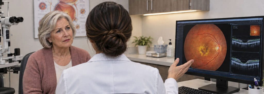
Small drusen are common with age and relatively harmless, but larger or more numerous drusen are a hallmark early warning sign of AMD. Over time, oxidative stress, chronic inflammation, and reduced blood flow to the retina accelerate this cellular damage, ultimately impairing your central vision.
These risk factors are known to increase the likelihood of developing AMD:
Having a family history of AMD
Age (most cases are diagnosed after age 55)
Smoking (studies show that smoking is associated with 25% of severe AMD cases, and living with a smoker doubled the test subjects’ risk of developing AMD)
Obesity
Being Caucasian
Being female
Taking certain medications like chloroquine or phenothiazine-type drugs
Smoking is particularly damaging because it directly accelerates oxidative stress in the retinal tissue and reduces the oxygen supply to the macula, compounding the cellular breakdown already underway. Similarly, obesity and poor cardiovascular health can restrict blood flow to the eye, depriving the RPE of the nutrients it needs to function. While you cannot change your genetics or your age, addressing modifiable risk factors early is one of the most meaningful steps you can take to protect your long-term vision.
What are the symptoms of macular degeneration?
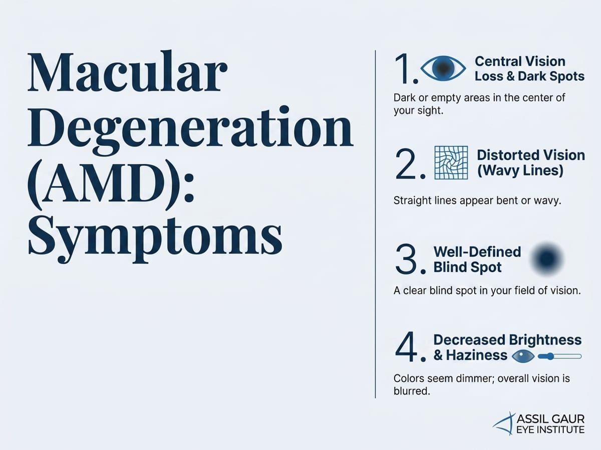
Macular degeneration affects the macula, the small central region of the retina responsible for sharp, detailed vision. Because it targets central rather than peripheral vision, the symptoms can be subtle at first and easy to dismiss as normal aging. Knowing what to watch for is the first step toward protecting your sight.
Loss of central vision or dark spots in the center of vision
A well-defined blind spot in your field of vision
Distorted vision, with straight lines appearing wavy
Decreased brightness of colors and overall haziness in your vision
Difficulty adjusting to changes in lighting
Needing more light to perform tasks
Loss of depth perception
Blurred vision
One of the most practical tools for catching early changes at home is the Amsler grid, a simple checkerboard pattern with a dot at the center. When you cover one eye and focus on the center dot, any wavy, blurry, or missing areas in the grid lines can signal that the macula is being affected. Many patients are surprised to discover a difference between their two eyes when they test them separately. Your care team at Assil Gaur Eye Institute can provide you with an Amsler grid and walk you through using it as part of your ongoing monitoring routine.
Symptoms can also vary significantly between the two main types of the disease. Dry AMD tends to progress slowly over months or years, with gradual blurring and a growing need for brighter light. Wet AMD, by contrast, can cause rapid and severe central vision loss over days or even hours as abnormal blood vessels leak fluid beneath the retina.
When to seek urgent care: If you notice a sudden increase in distortion, a new dark spot in your central vision, or any rapid change in how clearly you can see, treat it as an urgent medical issue and contact our team right away. Wet AMD responds best to treatment when caught early, and prompt action can make a meaningful difference in preserving your vision. Trusted by patients across Los Angeles and beyond, our world-class retinal specialists can evaluate and act quickly when it matters most.
Is there a cure for macular degeneration?
No. However, the last decade has made significant breakthroughs in treatment options. Today, some medications help control and slow AMD progression and, in some cases, may even improve vision.
What are the stages of macular degeneration?
Understanding how macular degeneration progresses can help you and your doctor make smarter decisions about monitoring and treatment. The staging system applies primarily to dry AMD, which accounts for the vast majority of cases. Doctors generally recognize three stages.
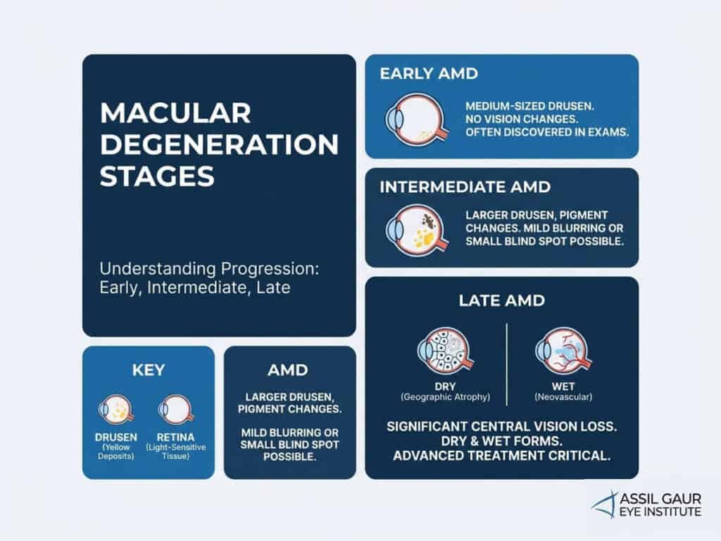
Early AMD
Early AMD is characterized by medium-sized drusen, tiny yellow deposits that accumulate beneath the retina. At this stage, most people experience no noticeable vision changes. Because there are no obvious symptoms, people often discover early AMD only during a routine dilated eye exam, which is one of the strongest reasons to keep up with regular comprehensive eye care. Read more about protecting your vision proactively in our guide to the 10 commandments for healthy eyes.
Intermediate AMD
Intermediate AMD involves larger drusen, pigment changes in the retina, or both. Some people begin to notice mild blurring or a small blind spot near the center of their vision, though many still have no symptoms. This is the stage where lifestyle modifications and nutritional supplements may help slow progression.
Late AMD
Late AMD involves significant central vision loss. Late dry AMD, sometimes called geographic atrophy, involves the breakdown of light-sensitive cells in the macula. Late wet AMD develops when abnormal blood vessels grow beneath the retina and leak fluid, causing rapid and severe vision changes. At this stage, advanced treatment becomes critical.
Knowing your stage empowers you to take the right steps at the right time, and world-class monitoring makes all the difference in catching progression early.
There are two types of macular degeneration. How are they different?
The two forms of macular degeneration are dry and wet. Both affect the macula, part of the retina. They differ in how quickly they develop and how severe the vision loss can be.
Dry AMD is the more common form, accounting for about 80% of all cases. It develops gradually over years, progressing through early, intermediate, and late stages as tiny deposits called drusen accumulate beneath the retina. Wet AMD, by contrast, affects roughly 20% of dry AMD patients who progress to it and is far more aggressive. It can develop over weeks to months and carries a much higher risk of rapid, irreversible central vision loss if left untreated.
The key distinction comes down to what is happening inside the eye. In dry AMD, retinal cells slowly break down as metabolic waste builds up. In wet AMD, the body responds by producing excess Vascular Endothelial Growth Factor (VEGF), a protein that triggers the growth of fragile, leaking blood vessels beneath the macula. That leakage is what makes wet AMD so urgent to treat.
Both forms require expert monitoring and care. At Assil Gaur Eye Institute, our retinal specialist, Dr. Svetlana Pilyugina, brings world-class precision to diagnosing and managing macular degeneration. As one of only approximately 3,000 vitreoretinal specialists in the country, Dr. Pilyugina offers patients the reassurance of truly elite, advanced care from the very first evaluation.
Read on to understand each form in detail, how they are diagnosed, and what treatment options are available to protect your vision.
What is dry macular degeneration?
Dry AMD is the less severe early stage and is more common. Eighty percent of AMD patients have a dry form, which gradually worsens over the years.
In the dry stage, tiny yellow deposits of metabolic waste, known as ¨drusen¨, form in the macula. At first, they have little effect on vision. However, as the disease progresses and drusen accumulate, they obstruct the flow of nutrients and oxygen to the macula.
How is dry macular degeneration treated?
Valeda Light Delivery System
The Valeda Light Delivery System is a non-invasive, FDA-authorized treatment for dry age-related macular degeneration (AMD) that uses targeted light therapy to support retinal cell health. Instead of injections or surgery, Valeda delivers specific wavelengths of red, amber, and near-infrared light to the retina-a process called photobiomodulation. This light stimulates the mitochondria within retinal cells, helping them produce more energy, reduce inflammation, and function more effectively over time.
Treatment is performed in-office and typically involves a series of short sessions over several weeks, with no downtime or recovery required. While it does not cure AMD, clinical studies show that patients experience improved vision and slowed disease progression, particularly in those with early to intermediate stages.
AREDS2
One established treatment for dry AMD is the AREDS2 formula, a combination of antioxidant supplements and key visual pigments (lutein and zeaxanthin) that support retinal health. While it is not a cure, AREDS2 has been shown to reduce the risk of progression to advanced (wet) AMD by approximately 25% in appropriate patients.
What is wet macular degeneration?
Wet macular degeneration (Wet AMD) is the more advanced form of this disease, and about 20% of Dry AMD patients will progress to it. Also, if the condition has appeared quickly over weeks to months, it is also wet AMD.
The wet stage of AMD is aggressive and requires urgent treatment by a retinal specialist to prevent progressive vision loss. If wet AMD goes untreated, blood or fluid buildup in the retina leads to scarring, which can cause irreversible vision loss.
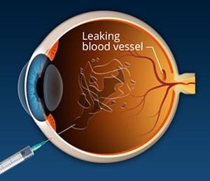
Eyes with wet AMD produce more vascular endothelial growth factor (VEGF). This protein triggers the growth of new blood vessels in the retina. Too much VEGF in the eye creates fragile, malformed blood vessels. These vessels leak, damaging the macula and surrounding retina and eventually leading to central vision loss.
If you get wet AMD in one eye, you’ll have a 20% chance of developing it in your other eye within five years.
How is wet macular degeneration treated?
Once AMD progresses to the wet form, pills are no longer an acceptable treatment option. Instead, doctors must place medications directly in the eye to control the disease at its source. Luckily, injectable medications have revolutionized the treatment for wet AMD.
Although they sound scary, intraocular injections are quick, painless, safe, and effective in slowing disease progression – and in some cases, restoring some of the lost vision. These medications target a protein called vascular endothelial growth factor, or VEGF. In wet AMD, abnormal blood vessels beneath the retina leak fluid and blood, causing rapid central vision loss. Anti-VEGF medications block this protein, stopping the growth of those leaky vessels and allowing the retina to stabilize.
Several advanced anti-VEGF agents are available today. Lucentis (ranibizumab) and Eylea (aflibercept) have decades of clinical evidence behind them and remain trusted cornerstones of wet AMD care. Newer agents including Beovu (brolucizumab) and Vabysmo (faricimab) offer the potential for longer intervals between injections, which can meaningfully reduce the treatment burden for patients who require ongoing care. Vabysmo, in particular, targets two pathways simultaneously, giving our retinal specialist additional precision in tailoring your treatment plan.
Most patients begin treatment with monthly injections until the disease stabilizes, after which dosing frequency is often extended to every two, three, or even four months depending on how the eye responds. Our world-class retinal specialist, Dr. Svetlana Pilyugina, closely monitors each patient using advanced imaging to determine the ideal interval, ensuring you receive exactly the level of care your vision requires.
In some cases, a laser treatment called photodynamic therapy may also be effective. Dr. Pilyugina will recommend the option, or combination of options, best suited to your individual situation.
Living with macular degeneration: tips for daily life
A macular degeneration diagnosis can feel overwhelming, but many people continue to live full, independent lives with the right strategies in place. Because AMD affects central vision rather than peripheral vision, you can adapt your environment and daily routines to stay safe and confident at home.
Here are some practical tips that can make a real difference:
Maximize your lighting. Bright, even lighting reduces strain when you have reduced central vision. Replace dim bulbs throughout your home, especially in reading areas and the kitchen, and consider adjustable task lamps for close-up work.
Use magnification tools. Handheld magnifiers, digital magnifying glasses, and screen-enlarging software on phones and computers can help you read, manage medications, and handle everyday tasks more easily.
Increase contrast. High-contrast settings on digital devices, large-print books, and bold-lined writing paper all make text easier to distinguish when central vision is compromised.
Lean on your peripheral vision. A low-vision specialist can teach you eccentric viewing techniques that train you to use the peripheral parts of your visual field more effectively for reading and recognizing faces.
Prioritize eye-healthy nutrition. Leafy greens, colorful vegetables, and omega-3-rich foods support overall retinal health. Our guide on whether green smoothies can support eye health is a great place to start.
Monitor changes with an Amsler grid. Keeping one on your refrigerator and checking it daily helps you catch any sudden shifts in your vision quickly so your care team can respond promptly.
Working closely with a world-class retinal specialist gives you the best foundation for managing macular degeneration with confidence and preserving your vision for as long as possible.
Can you prevent macular degeneration?
No, but simple lifestyle changes can meaningfully decrease your risk of developing it. Diet plays a bigger role than most people realize. Eating plenty of dark green leafy vegetables like kale, spinach, and collard greens gives your eyes lutein and zeaxanthin, two nutrients that help protect the macula from oxidative damage. Eggs, corn, and orange bell peppers are also excellent sources of these same protective carotenoids. Salmon and other cold-water fish supply the omega-3 fatty acids your retinal cells depend on to stay healthy.
If you have intermediate AMD or advanced AMD in one eye, your doctor may recommend an AREDS2 supplement formula. Clinical research has shown that this specific combination of vitamins C and E, lutein, zeaxanthin, zinc, and copper can reduce the risk of progression to advanced AMD by about 25 percent. It is worth asking your specialist whether AREDS2 supplementation makes sense for your stage of the disease.
It is also essential to exercise regularly and keep both blood pressure and cholesterol under control. Chronically elevated blood pressure restricts blood flow to the retina and accelerates damage to the delicate vessels that feed the macula, so working with your primary care physician to reach healthy targets is a genuinely important part of protecting your vision in the long term. Wear sunglasses that provide 100% UV protection and shield your eyes from visible light and blue light. Lastly, if you smoke, stop. Research consistently shows that smokers face roughly double the risk of developing AMD compared to non-smokers.
If AMD runs in your family, consider asking about genetic testing. Variants in genes like CFH and ARMS2 are strongly associated with elevated risk, and knowing your genetic profile allows your care team to personalize your monitoring schedule and prevention strategy before any vision loss begins.
We cannot overstate the importance of getting an annual eye exam to screen for AMD and other eye health conditions that can threaten your vision and quality of life.
Call now or book your appointment online.
How is macular degeneration diagnosed?
Even if you have no noticeable symptoms yet, routine eye exams are one of the most powerful tools for catching macular degeneration early. Because the early stages of AMD can be completely silent, a comprehensive evaluation by a world-class retinal specialist like Dr. Svetlana Pilyugina is essential for anyone over 50 or with known risk factors. Here are the key diagnostic tests used to detect and monitor AMD.
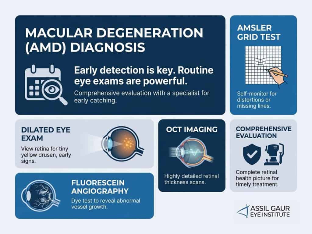
Amsler grid test. This simple but effective test involves looking at a grid of straight lines. If the lines appear wavy, blurry, or distorted, or if sections of the grid seem to disappear, that can be an early warning sign of macular changes. It is also a test patients can perform at home between appointments to monitor for any shifts in their vision.
Dilated eye exam. Your doctor will use dilating drops to widen your pupils, allowing a clear view of the retina and macula. This is where drusen, the tiny yellow deposits that signal early AMD, are typically first detected.
Optical coherence tomography (OCT). OCT uses light waves to produce highly detailed, cross-sectional images of the retina. It allows your specialist to measure retinal thickness and detect fluid buildup or structural changes with remarkable precision, even before symptoms appear.
Fluorescein angiography. A special dye is injected into a vein in your arm, and as it travels through the blood vessels of the retina, a camera captures images that reveal leaking, abnormal blood vessel growth, or areas of poor circulation, all hallmarks of wet AMD.
Together, these advanced diagnostic tools give your care team a complete picture of your retinal health so treatment can begin at exactly the right moment.
What is the outlook for people with macular degeneration?
One of the first questions people ask after a macular degeneration diagnosis is a simple one: what does my future look like? The honest answer is that it depends on the type and stage of the disease, but there is genuine reason for optimism, especially when the condition is caught and managed early.
The most important thing to understand is that macular degeneration, even in advanced stages, does not cause complete blindness. The disease affects your central vision, but your peripheral vision typically remains intact. Most people with macular degeneration retain enough functional vision to stay independent and continue enjoying daily activities with the right support and low-vision tools.
For dry AMD, progression is usually slow. Many people spend years in the early or intermediate stage without noticing significant changes to their vision. Adopting healthy habits, including a nutrient-rich diet and consistent monitoring, can meaningfully slow that progression.
Wet AMD moves faster, but it also responds well to treatment. Anti-VEGF injections have transformed the outlook for wet AMD patients over the past two decades, with many people stabilizing their vision and some even experiencing meaningful improvement. Advanced therapies like the Valeda Light Delivery System are further expanding those possibilities.
The single biggest factor in a positive outcome is consistent, world-class care from a specialist who knows your eyes. Early detection, regular monitoring, and access to the latest treatments give you the best possible chance at preserving the vision you have for years to come.
Experience Assil Gaur Eye Institute’s nationally recognized macular degeneration care.
The AGEI staff includes a highly skilled Stanford-trained retina specialist, Dr. Svetlana Pilyugina (known to her patients as “Dr. P”). Dr. P is an ophthalmologist with fellowship training and board certification in diseases and surgery of the vitreous and retina.
Dr. Pilyugina has extensive experience treating macular degeneration, retinal detachment, diabetic retinopathy, and macular holes and puckers, having treated thousands of patients over the past decade.
As a member of an elite group of only 3000 vitreoretinal specialists in the United States, Dr. Pilyugina brings a unique skill set to AGEI in treating and surgically treating retinal disease. Her academic credentials include numerous research papers, conference presentations, medical publications, and ophthalmology clinical trials.
We are conveniently located for patients throughout Southern California and the Los Angeles area, including Beverly Hills, Santa Monica, West Los Angeles, West Hollywood, Culver City, Hollywood, Venice, Marina del Rey, Malibu, Manhattan Beach, and Downtown Los Angeles.
Read real patient stories about retinal conditions.
MACULAR DEGENERATION FAQs
Is macular degeneration hereditary?
Yes, macular degeneration can have a hereditary component. However, a genetic predisposition does not necessarily mean an individual will develop macular degeneration. Lifestyle factors such as smoking, diet, and sun exposure can also increase the risk of developing the disease.
What are the early warning signs of macular degeneration?
The early warning signs of macular degeneration may include:
- Objects appear distorted or blurry, or straight lines look wavy.
- Needing help seeing in dimly lit environments, such as restaurants or movie theaters.
- Colors appear less vivid, or it is difficult to distinguish between shades of colors, and you cannot see fine details.
- A dark spot in the center of your visual field.
- Prolonged adaptation to changes in lighting.
Can macular degeneration be reversed?
No, it cannot be reversed. However, early detection and treatment can slow or pause disease progression, allowing you to maintain your lifestyle during treatment.
What does vision look like with macular degeneration?
Macular degeneration can cause blurry or distorted vision in the center of the visual field. Usually, you’re still able to use your peripheral vision. You can learn to use your peripheral vision to do things with the help of a vision rehabilitation specialist who can teach you how it works.
What is the best eye vitamin for macular degeneration?
No specific vitamin or nutritional supplement has been proven to cure or prevent macular degeneration. However, a large National Eye Institute study called AREDS (Age-Related Eye Disease Study) found that certain vitamins and nutrients, like beta-carotene and vitamin E, may protect the eyes in some people.
Which is more severe wet or dry macular degeneration?
Wet (exudative) macular degeneration is generally considered more severe and can progress faster than dry (atrophic) macular degeneration.
How fast does macular degeneration progress?
In general, dry macular degeneration can take years or even decades to progress to the more advanced stages, where central vision loss becomes more noticeable.
Wet macular degeneration can progress rapidly and cause severe vision loss in weeks or months if left untreated.
How is macular degeneration diagnosed?
Macular degeneration is typically diagnosed through a comprehensive eye exam that may include the following tests:
- Visual acuity test: Using an eye chart, this test measures how well you can see at different distances.
- Dilated eye exam: During this exam, your eye care provider will use dilating eye drops to widen your pupils and examine the back of your eye, including the macula, for signs of macular degeneration and other eye conditions.
- An Amsler grid test allows the patient to look at a grid, identify all four corners, and the doctor to analyze the response for distortion or missing areas in the central vision.
- Optical coherence tomography (OCT): This non-invasive imaging test uses light waves to create a detailed cross-sectional image of the retina, allowing your eye doctor to see any changes or damage to the macula.
- Fluorescein angiography: In this test, a dye is injected into a vein in your arm. Your eye doctor will take photographs of your retina as the dye circulates through your blood vessels. This can help identify abnormal blood vessels or leakage causing wet macular degeneration.
What foods should be avoided with macular degeneration?
Foods that should be avoided or limited include:
- Processed and fried foods: These foods are high in unhealthy fats, contributing to inflammation and increasing the risk of macular degeneration.
- Sugary and high-hypoglycemic-index foods: These foods can cause spikes in blood sugar levels, contributing to inflammation and increasing the risk of macular degeneration.
- Red and processed meats: These foods are high in saturated fats and can contribute to inflammation and increase the risk of macular degeneration.
- Trans fats: These are found in many processed and baked goods, contributing to inflammation and increasing the risk of macular degeneration.
What percentage of macular degeneration patients go blind?
Most people with macular degeneration do not go completely blind. Still, they may experience significant low vision, making it difficult or virtually impossible to perform everyday activities like reading, driving, and recognizing faces.
Can you have both glaucoma and macular degeneration?
Yes, a person can have both glaucoma and macular degeneration. Although these are two different eye conditions, they can coincide, especially in older adults.
What is the difference between diabetic macular edema and macular degeneration?
Both conditions affect the macula and can impair central vision, but their causes differ. Diabetic macular edema results from diabetes-related retinal swelling, while macular degeneration is typically associated with aging. Our retina specialist can diagnose the condition and recommend the most effective treatment plan.
Are flashes and floaters a sign of macular degeneration?
-
AREDS 2 Supplements for Age-Related Macular Degeneration (AMD) | National Eye Institute
-
Age-Related Macular Degeneration (AMD) | National Eye Institute
-
Vision Loss, Central – American Academy of Ophthalmology
-
Ocular nutritional supplements: are their ingredients and manufacturers’ claims evidence-based?
https://pubmed.ncbi.nlm.nih.gov/25458196 -
Dietary fatty acid intake, plasma fatty acid levels, and the risk of age-related macular degeneration (AMD): a dose-response meta-analysis of prospective cohort studies
-
Macular degeneration: peculiar sunlight exposure in an agricultural worker
-
Omega-3 fatty acids for preventing or slowing the progression of age‐related macular degeneration
https://www.ncbi.nlm.nih.gov/pmc/articles/PMC7087473/

Written by The AGEI Educational Team

Medically Reviewed by Dr. Svetlana Pilyugina
No form shortcode found.