Macular Holes & Macular Puckers in Los Angeles
in Los Angeles
Understanding the Macula
The macula is a part of the eye located in the center of the retina, the light-sensitive tissue at the back of the eye. It’s a small, sensitive area that provides central vision and enables one to see fine details clearly.
The macula is critical for tasks such as reading, driving, and recognizing faces. This region also contains a high concentration of cone cells, which are responsible for color vision
Dr. Pilyugina talks about macular holes
What’s a macular pucker?
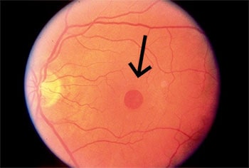
A macular pucker, also known as an epiretinal membrane (ERM), is a condition where a thin layer of scar tissue forms on the surface of the macula. This scar tissue can cause the macula to wrinkle or pucker up, resulting in distorted and blurred vision.
Macular puckers can develop after eye trauma, retinal detachment, or in patients with inflammatory conditions. Most commonly, they happen in otherwise healthy eyes. In most cases, it’s due to changes in the eye as we age.
What is a macular hole?
A macular hole commonly affects people over the age of 55 and often occurs in females. Most macular holes develop spontaneously, without a known cause. For this reason, there is no effective way to prevent their formation.
A macular pucker involves tissue forming on the macula, while a macular hole is a hole or tear in the macula. Macular holes can seriously affect your ability to drive, read, and do other precision tasks.
Like macular puckers, macular holes can occur as the eye changes with age.
The risk of developing a macular hole is increased for:
Persons who have had blunt eye trauma
Those with high myopia (nearsightedness)
People with diabetes or those with diabetic eye disease
Macular pucker: formation of a scar tissue layer over the macula can become distorted and bent, causing the retina to wrinkle.
Vitreous traction: the vitreous is a “gel” that fills up the inside of the eye.
Aging, the vitreous can shrink and pull away from the retina, occasionally causing a macular hole.
Individuals who have already had a macular hole have a 5% to 15% risk of developing one in their other eye.
What are the symptoms of a macular pucker and a macular hole?
The most common symptoms include:
A gradual decline in central vision.
Blurred or distorted vision (where straight lines will appear wavy)
A growing dark spot in the central visual field.
Diagnosing a macular hole or pucker requires a dilated eye exam with a close inspection of the retina. At AGEI, we use the industry gold standard for diagnostic testing: optical coherence tomography (OCT).
This non-invasive ocular imaging is quick and painless. It allows high-resolution evaluation of the macula using reflected light. It enables our specialists to differentiate, diagnose, and manage macular holes and other eye conditions.
Treatment for Macular Holes and Macular Puckers
Treatment depends on severity. For minor cases, our specialist may recommend regular check-ups to ensure the condition is not worsening or causing further complications.
In some cases, surgical intervention may be needed to prevent further damage and preserve vision.
Macular pucker surgery
Eye drops, medications, or supplements cannot improve vision caused by an epiretinal membrane. The only treatment option is surgery. If a macular pucker affects your vision, doctors may perform a vitrectomy. This procedure involves removing the vitreous gel (a gel-like substance inside the eye that helps keep it round) and peeling off scar tissue from the retina’s surface.
Macular hole eye surgery
A vitrectomy can also treat a macular hole. In this case, once the vitreous gel is removed, the eye is filled with a special gas bubble that helps flatten the edges of the macular hole against the eyewall, allowing it to grow back together.
Experience Assil Gaur Eye Institute’s nationally recognized expertise in treating macular holes and macular puckers
The AGEI ophthalmology group includes a highly skilled retina specialist, Dr. Svetlana Pilyugina, or “Dr. P,” as her patients know her. Dr. Pilyugina is an ophthalmologist with fellowship training and board certification in vitreoretinal diseases and surgery.
Dr. P has extensive experience treating retinal diseases, including age-related macular degeneration, diabetic eye disease and diabetic retinopathy, retinal tears and detachment, flashes, floaters, macular holes, retinal vein occlusion, posterior vitreous detachment, and other complex retinal conditions.
In addition to her medical experience, Dr. P is known for her dedication to her patients and focus on personal service. She’s committed to dedicating each day to her patients and providing the highest standards of personalized care.
We are conveniently located for eye care patients throughout Southern California and the Los Angeles area, including Beverly Hills, Santa Monica, West Los Angeles, West Hollywood, Culver City, Hollywood, Venice, Marina del Rey, Malibu, Manhattan Beach, and Downtown Los Angeles.
Assil Gaur Eye Institute has extensive experience in a full range of vision problems, including glaucoma, cataract surgery, LASIK eye surgery, and retinal diseases, to name just a few.
Read real patient stories about retinal conditions.
Call now or book your appointment online.
Macular pucker and macular hole FAQs
How successful is Vitrectomy Surgery for ERM?
Epiretinal membrane peels have a very good success rate, and most patients achieve improved visual acuity with reduced distortion.
ERM develops over many months; it takes time (several months) for the retinal surface tissue to relax and resume its normal anatomic shape after surgery. Depending on the size and age of the ERM, restoring vision entirely may not be possible.
What’s macular hole surgery recovery like?
The surgery performed to repair the macular hole is unique because a gas bubble is placed in the eye. It’s important to follow our specialist postoperative instructions to ensure the gas bubble remains in contact with the macula following surgery. This will allow the macular hole to stay pressed against the eyewall.
The patient must lie face down throughout the day and continue this routine for up to 14 days postoperatively. This is necessary because the gas rises, and the only way to keep the bubble in contact with the macula is to keep the head face down. This is extremely important to the success of the surgery since, with proper positioning, the hole has a better chance of healing.
The gas bubble will slowly dissolve and shrink over time. While gas is present in the eye, it is critical to avoid air travel until the surgeon clears you to do so. Gases expand at altitude and can cause an extreme elevation in ocular pressure, leading to irreversible vision loss.
What is a “full-thickness” macular hole?
A macular hole is considered “full thickness” when it extends through the entire retina, from the internal limiting membrane on the surface to the photoreceptor layer at the bottom. This typically occurs at stage 2 or later in macular hole progression. These usually won’t heal on their own, and macular hole closure surgery may be required.
What’s the closure rate of a macular hole?
The success rate of surgically closing a macular hole using vitrectomy surgery is as high as 90-95% after a single surgery.
Why is a retinal surgeon the best doctor for macular hole repair?
A retinal surgeon specializes in diseases and conditions that affect the retina, including macular holes. They have extensive training and experience diagnosing and treating these specific eye problems.
Repairing a macular hole is a delicate surgical procedure known as a vitrectomy. This surgery is intricate and requires precise manipulation of the retina, skills that our retinal surgeons have developed through years of specialized training.
Is there a natural treatment for macular pucker?
No natural remedy exists for conditions like a macular hole or a macular pucker, also known as an epiretinal membrane. The only scientifically proven method to treat this issue is through surgical intervention, which we typically reserve for more severe cases.
Is macular pucker surgery an outpatient procedure?
Yes, macular pucker surgery, commonly known as a vitrectomy, is typically an outpatient procedure.
Can a macular hole heal on its own?
In rare cases, a small macular hole (Stage 1) may close spontaneously without treatment. However, most macular holes do not heal on their own and will progress without surgical intervention. Early diagnosis is critical — the sooner a macular hole is identified and treated, the better the chance of restoring visual acuity. If you notice any sudden change in your central vision, contact a fellowship-trained retina specialist promptly.
How long does recovery take after macular hole surgery?
Recovery after vitreoretinal surgery for a macular hole typically takes several weeks to months. One of the most important aspects of recovery is face-down positioning: after the gas bubble is placed inside the eye, patients must remain face-down for days to weeks so the bubble can press against the macular hole and help it close. Vision improves gradually, and most patients see meaningful gains within 4 to 8 weeks as the gas bubble is slowly absorbed by the eye.
What is the success rate of macular hole surgery?
Vitrectomy for macular holes has a closure rate of over 90%, making it one of the most effective procedures in vitreoretinal surgery. At Assil Gaur Eye Institute, our world-class retina specialist brings fellowship-trained expertise and advanced surgical precision to every procedure, giving Los Angeles patients the best possible chance of a full recovery.
Can a macular hole or macular pucker affect both eyes?
Yes. People with a macular hole in one eye have a 5% to 15% risk of developing one in the other eye. Macular puckers can also develop bilaterally, though this is less common. Regular dilated eye exams are essential to monitor both eyes and catch any changes in the macula early.
What is the difference between a macular hole and a macular pucker?
A macular pucker (epiretinal membrane) is a thin layer of scar tissue that forms on top of the macula, causing it to wrinkle and distort vision. A macular hole is an actual break or opening through the macula, which can cause a dark spot or significant loss of central vision. Both conditions are diagnosed using optical coherence tomography (OCT) and, when treatment is needed, are addressed through vitrectomy surgery.
-
Dynamics of macular hole closure in gas-filled eyes within 24 h of surgery observed with swept-source optical coherence tomography.
https://karger.com/ore/article-abstract/53/1/48/258553/Dynamics-of-Macular-Hole-Closure-in-Gas-Filled?redirectedFrom=fulltext -
Morpho-functional evaluation of full-thickness macular holes by the integration of optical coherence tomography angiography and microperimetry.
https://doi.org/10.3390/jcm9010229 -
hVitreous surgery for idiopathic macular holes: results of a pilot study.
https://doi.org/10.1001/archopht.1991.01080050068031

Written by The AGEI Educational Team

Medically Reviewed by Dr. Svetlana Pilyugina
No form shortcode found.