What is the macula?
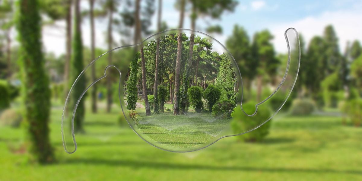
The macula is a part of the eye located in the center of the retina, the light-sensitive tissue at the back of the eye. It's a small, sensitive area that provides central vision and enables one to see fine details clearly.
The macula is critical for tasks such as reading, driving, and recognizing faces. This region also contains a high concentration of cone cells, which are responsible for color vision
- What's a macular pucker?
- What is a macular hole?
- What are the symptoms of a macular pucker and macular hole?
- Treatment for Macular holes and macular puckers
- Experience Assil Gaur Eye Institute's nationally recognized expertise in treating macular holes and macular puckers
- Macular pucker and macular hole FAQs
- How successful is Vitrectomy Surgery for ERM?
- What's macular hole surgery recovery like?
- What is a “full-thickness” macular hole?
- What’s the closure rate of a macular hole?
- Why is a retinal surgeon the best doctor for macular hole repair?
- Is there a natural treatment for macular pucker?
- Is macular pucker surgery an outpatient procedure?
What's a macular pucker?

A macular pucker, also known as an epiretinal membrane (ERM), is a condition where a thin layer of scar tissue forms on the surface of the macula. This scar tissue can cause the macula to wrinkle or pucker up, resulting in distorted and blurred vision.
Macular puckers can develop after eye trauma, a retinal detachment, or in patients with inflammatory conditions. But most commonly, they happen in otherwise completely healthy eyes. In most cases, it’s due to changes in the eye as we age.
What is a macular hole?
A macular hole commonly affects people over the age of 55 and often occurs in females. The vast majority of macular holes develop spontaneously and without cause. It is for this reason that there is no way to prevent their formation or development effectively.
While a macular pucker involves tissue forming on the macula, a macular hole is a hole or tear in the macula. Macular holes can seriously affect your ability to drive, read, and do other precision tasks.
Much like macular puckers, macular holes occur due to changes in the eye as we age.
The risk of developing a macular hole is increased for:
- Persons who have had blunt eye trauma
- Those with high myopia (nearsightedness)
- Diabetics or those with diabetic eye disease
- Macular pucker: formation of a scar tissue layer over the macula can become distorted and bent, causing the retina to wrinkle.
- Vitreous traction: the vitreous is a “gel” that fills up the inside of the eye.
- Aging, the vitreous can shrink and pull away from the retina, occasionally causing a macular hole.
Individuals who have already suffered a macular hole have a 5% to 15% risk of developing a macular hole in their other eye.
Schedule your consultation today with our internationally recognized retina specialists
What are the symptoms of a macular pucker and macular hole?
The most common symptoms include:
- A gradual decline in central vision.
- Blurred or distorted vision (where straight lines will appear wavy)
- A growing dark spot in the central visual field.
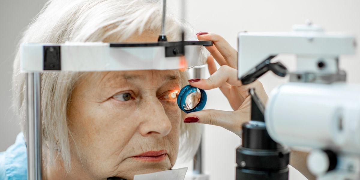
Diagnosing a macular hole or pucker requires a dilated eye exam with a close inspection of the retina. At AGEI we only use the Gold Standard in the industry for diagnostic testing and diagnosis called Optical coherence tomography (OCT).
This non-invasive ocular imaging is quick and painless. It allows for the evaluation of the macula in high resolution using reflected light and allows our specialist to differentiate, diagnose, and manage macula holes from other eye conditions and diseases.
Treatment for Macular holes and macular puckers
Treatments are determined based on the severity. For minor cases, our specialist may opt for regular check-ups, ensuring that the condition is not deteriorating or causing further complications.
Or surgical intervention may be needed in some instances to avoid additional damage and preserve vision.
Macular pucker surgery
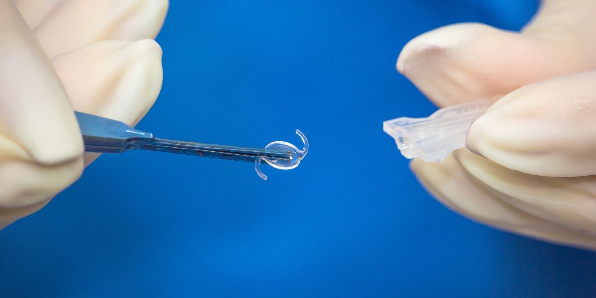
Eye drops, medications, or supplements cannot improve vision caused by an epiretinal membrane. The only treatment option is surgery. If your vision is affected by a macular pucker, a vitrectomy may be performed. This procedure involves removing the vitreous gel (gel-like substance inside the eye that helps keep its round shape) and peeling off scar tissue from the surface of the retina.
Macular hole eye surgery
A vitrectomy can also be performed to treat a macular hole. In this case, once the vitreous gel is removed, the eye is filled with a special gas bubble that helps to flatten the edges of the macular hole against the eyewall to allow it to grow back together.
Experience Assil Gaur Eye Institute's nationally recognized expertise in treating macular holes and macular puckers
The AGEI ophthalmology group includes a highly skilled retina specialist, Dr. Svetlana Pilyugina, or “Dr. P,” as she is known to her patients. Dr. Pilyugina is an ophthalmologist with fellowship training and board certification in diseases and surgery of the vitreous and retina.
Dr. P has extensive experience treating retinal diseases, including age-related macular degeneration, diabetic eye disease and diabetic retinopathy, retinal tears and detachment, flashes, floaters, macular holes, retinal vein occlusion, posterior vitreous detachment, and other complex retinal conditions.
In addition to her medical experience, Dr. P is known for her dedication to her patients and focus on personal service. She’s committed to dedicating each day to her patients, offering the highest standards of personalized care.
We are conveniently located for eye care patients throughout Southern California and the Los Angeles area in or near Beverly Hills, Santa Monica, West Los Angeles, West Hollywood, Culver City, Hollywood, Venice, Marina del Rey, Malibu, Manhattan Beach, and Downtown Los Angeles.
Assil Gaur Eye Institute has extensive experience in a full range of vision problems, including glaucoma, cataract surgery, LASIK eye surgery, and retinal diseases, to name just a few.
Macular pucker and macular hole FAQs
How successful is Vitrectomy Surgery for ERM?
Epiretinal membrane peels have a very good success rate, and most patients achieve improved visual acuity with reduced distortion.
ERM develops over many months, it takes time (several months) for the retinal surface tissue to relax and resume its normal anatomic shape after surgery. Depending on the size and age of the ERM, in some cases restoring vision completely is not possible.
What's macular hole surgery recovery like?
The surgery performed to repair the macular hole is unique because a gas bubble is placed in the eye. It’s important to follow our specialist post-operative instructions to ensure the gas bubble remains in contact with the macula following surgery. This will allow the macular hole to remain pressed against the eyewall.
The patient must lie face down throughout the day and continue this routine for up to 14 days postoperative. This is necessary because the gas rises, and the only way to keep the bubble in contact with the macula is to keep the head in a face-down position. This is extremely important to the success of the surgery since, with proper positioning, the hole has a better chance of healing.
The gas bubble will slowly dissolve and shrink over time. While gas is present in the eye, it is critical to avoid airplane or altitude travel until cleared to do so by the surgeon. Gases expand at altitudes and can cause an extreme elevation in ocular pressure, leading to irreversible vision loss.
What is a “full-thickness” macular hole?
A macular hole is considered "full thickness" when it goes through the entire thickness of the retina, from the internal limiting membrane on the surface to the photoreceptor layer at the bottom. This typically occurs at stage 2 and beyond in the progression of a macular hole. These normally won't heal on their own, and macular hole closure surgery may be required.
What’s the closure rate of a macular hole?
The success rate of surgically closing a macular hole using vitrectomy surgery is as high as 90-95% after a single surgery.
Why is a retinal surgeon the best doctor for macular hole repair?
A retinal surgeon specializes in diseases and conditions that affect the retina, including macular holes. They have extensive training and experience in diagnosing and treating these specific types of eye problems.
Repairing a macular hole is a delicate surgical procedure known as a vitrectomy. This surgery is intricate and requires precise manipulation of the retina, skills that our retinal surgeons have developed through years of specialized training.
Is there a natural treatment for macular pucker?
No natural remedy is available for conditions like a macular hole or a macular pucker, also commonly known as an epiretinal membrane. The only scientifically proven method to treat this issue is through surgical intervention, which we typically reserve for more severe instances.
Is macular pucker surgery an outpatient procedure?
Yes, macular pucker surgery, commonly known as a vitrectomy, is typically an outpatient procedure.
Sources
Kikushima W, Imai A, Toriyama Y, Hirano T, Murata T, Ishibashi T. Dynamics of macular hole closure in gas-filled eyes within 24 h of surgery observed with swept-source optical coherence tomography. Ophthalmic Res. 2015;53:48–54. https://doi.org/10.1159/000368437.
Bacherini D, Savastano MC, Dragotto F, et al. Morpho-functional evaluation of full-thickness macular holes by the integration of optical coherence tomography angiography and microperimetry. J Clin Med. 2020;9:229. https://doi.org/10.3390/jcm9010229.
Kelly NE, Wendel RT. Vitreous surgery for idiopathic macular holes: results of a pilot study. Arch Ophthalmol. 1991;109:654–9. https://doi.org/10.1001/archopht.1991.01080050068031.
National Eye Institute. Macular Pucker. (https://www.nei.nih.gov/learn-about-eye-health/eye-conditions-and-diseases/macular-pucker...
-
Retina Specialist

As a member of an elite group of only 3000 retina-vitreous specialists in the United States, Stanford trained Dr. Pilyugina brings to AGEI a unique skill set in the treatment and surgery of retinal disease. Her academic credentials include numerous research papers, conference presentations, medical publications, and clinical trials.

 I was referred to Assil Eye institute by my sister and two nieces. I got my first appointment fairly quickly. All of the employ...Masoud D.
- Eyesight is critical and I always highly recommend you!David K.
- Strongly recommend the Assil Eye Institute. Their attention to detail, customer care, and the final result is and will be worth...Kyle H.
 Their whole staff is fabulous and so caring!!! The Annual Eye Exam that Dr May Performs is so extensive and done with such...Joan M.
- I am pleased with the service I have received. And I will continue to recommend to my friends and familyFelix C.
- The staff is personable and professional.Kurt E.
- Dr may is greatJennifer L.
- Very good treatment!Steven M.
- NopeCaroline M.
- Always too long of a waitCindra L.
- MD was very attentive to my concerns.Charles D.
- Everything was great, not much else I can sayBarbara M.
-
 Listen Now
What are Some Common Medications that can Cause or Worsen Glaucoma?
Read
Listen Now
What are Some Common Medications that can Cause or Worsen Glaucoma?
Read
-
 Listen Now
Patients are raving about the enVista® Envy™ Trifocal IOL Lens
Read
Listen Now
Patients are raving about the enVista® Envy™ Trifocal IOL Lens
Read
-
 Listen Now
Understanding Pinguecula: Causes, Symptoms, and Treatment
Read
Listen Now
Understanding Pinguecula: Causes, Symptoms, and Treatment
Read
-
 Listen Now
What Are the Causes of Double Vision and How Can They Be Addressed?
Read
Listen Now
What Are the Causes of Double Vision and How Can They Be Addressed?
Read
-
 Listen Now
The History of IOLs: You’ve Come A Long Way, Baby!
Read
Listen Now
The History of IOLs: You’ve Come A Long Way, Baby!
Read
-
 Listen Now
Can Vitamin D Help Relieve Dry Eye Disease? A New Study Says Yes!
Read
Listen Now
Can Vitamin D Help Relieve Dry Eye Disease? A New Study Says Yes!
Read
-
 Listen Now
Can Stress Increase Eye Pressure in Glaucoma Patients?
Read
Listen Now
Can Stress Increase Eye Pressure in Glaucoma Patients?
Read
-
 Valeda Light Delivery System for Dry Age-Related Macular Degeneration (AMD)
Read
Valeda Light Delivery System for Dry Age-Related Macular Degeneration (AMD)
Read
-
 Listen Now
Can Pupil Size Reveal Intelligence? Maybe Size Really Does Matter!
Read
Listen Now
Can Pupil Size Reveal Intelligence? Maybe Size Really Does Matter!
Read
-
 Listen Now
Cataract Lens Replacement Failure: What Can You Do About It?
Read
Listen Now
Cataract Lens Replacement Failure: What Can You Do About It?
Read
-
 Listen Now
Alcohol and Dry Eye Syndrome: Understanding the Connection
Read
Listen Now
Alcohol and Dry Eye Syndrome: Understanding the Connection
Read
-
 Kerry and Tanaz Assil focus on the fruits of their labor
Read
Kerry and Tanaz Assil focus on the fruits of their labor
Read
-
 How Autoimmune Diseases Affect Your Eyes: What You Need to Know
Read
How Autoimmune Diseases Affect Your Eyes: What You Need to Know
Read
-
 Breakthrough Eye Implant Technology Offers New Hope for Patients with Advanced Macular Degeneration
Read
Breakthrough Eye Implant Technology Offers New Hope for Patients with Advanced Macular Degeneration
Read
-
 Listen Now
Are Cataracts at a Young Age Getting More Common?
Read
Listen Now
Are Cataracts at a Young Age Getting More Common?
Read


Look Who Trusts Their Eyes to Assil Gaur Eye Institute...
-
LeBron James
Los Angeles LakersLeBron James is one of the NBA's greatest of all time. He absolutely depends on his vision to perform at the very highest level. That's why he's an AGEI LASIK surgery patient.
-
Anthony Davis
Dallas Mavericks -
Chris Paul
San Antonio Spurs -
Paul George
Philadelphia 76ers -
Marlee Matlin
Actress -
Michelle Williams
Actress -
Dwyane Wade
Miami Heat (retired) -
LaToya Jackson
Entertainer -
Lorenzo Lamas
Actor -
Philip Bailey
Earth Wind and Fire -
Gary Sinise
Actor -
Rip Hamilton
Former NBA player -
John Salley
Commentator, former NBA player -
Blair Underwood
Actor -
Tom ArnoldComedian/Actor
-
Troy EvansActor
-
John NobleActor
-
Isaac EddyActor/Director/Teacher
-
Barbara RushActress
-
John EmersonUS Ambassador to Germany
-
Shane MosleyFormer professional boxer
-
Cuttino MobleyFormer NBA player
-
Seth GordonDirector
-
Maurice EvansFormer NBA player
-
Wang LuoyongActor
-
Jesse MetcalfeActor
-
Ananda LewisTelevision Personality
-
Rob BrydonComedian
-
Clifton Collins Jr.Actor
-
Malinda WilliamsActress
-
Adrienne FrantzActress
-
Brad PittActor
-
LeBron JamesLos Angeles Lakers
-
Anthony DavisDallas Mavericks
-
Chris PaulSan Antonio Spurs
-
Courtney CoxActress
-
Paul GeorgePhiladelphia 76ers
-
Marlee MatlinActress
-
Michelle WilliamsActress
-
Dwyane WadeMiami Heat (retired)
-
Jalen BrunsonNew York Knicks
-
Mychal ThompsonCommentator, former NBA player
-
LaToya JacksonEntertainer
-
Lorenzo LamasActor
-
Philip BaileyEarth Wind and Fire
-
Gary SiniseActor
-
Rip HamiltonFormer NBA player
-
John SalleyCommentator, former NBA player
-
Blair UnderwoodActor
-
Tom ArnoldComedian/Actor
-
Troy EvansActor
-
John NobleActor
-
Isaac EddyActor/Director/Teacher
-
Barbara RushActress
-
Rudy GarciduenasBlue Man Group
-
John EmersonUS Ambassador to Germany
-
Shane MosleyFormer professional boxer
-
Cuttino MobleyFormer NBA player
-
Seth GordonDirector
-
Maurice EvansFormer NBA player
-
Cheri BlauwetPhysician and Wheelchair Racer
-
Wang LuoyongActor
-
Jesse MetcalfeActor
-
Jeana WilsonActress
-
Courtnee DraperActress
-
Ananda LewisTelevision Personality
-
Robyn JohnsonProducer
-
Rob BrydonComedian
-
Cary SchumanActor
-
Roland Buddy Lewis Jr.Comedian
-
Clifton Collins Jr.Actor
-
Malinda WilliamsActress
-
Chad JeffersMusician
-
Adrienne FrantzActress
-
Franco CarlottoBody Builder
-
Brad MatesMusician
-
David PichetteMusician
-
Mike MelaconProfessional Baseball Player
-
Nautica de la CruzRadio Personality
-
Lynn ConkwrightBody Builder
-
Nely GalanProducer
-
Roxanne GallaActress