- Home
- Resource Library
- Diagnostic Testing
- Retina Diagnostic Tests
Our State-of-the-Art Retina Diagnostic Tests and Tools

Assil Gaur Eye Institute's State-of-the-Art Retinal Diagnostic Testing Tools
When you visit a retina specialist, there are a number of tests that your doctor might do in order to diagnose or monitor certain retinal conditions. Because these tests aren’t typically performed at most optometrist offices, you may not be familiar with them. Don’t worry, they’re all painless and don’t involve poking your eye!
Here’s a quick list of retinal diagnostic tests you may experience at AGEI:
Digital Fundus Photography
This test uses specialized equipment to take a panoramic view of the inside of your eye in order to capture a detailed image of the retina, optic nerve, retinal blood vessels, and outer edges of the retina.
What to expect with digital fundus photography testing
Digital fundus photography is painless and non-invasive. After your eyes are dilated, you’ll be seated comfortably and asked to place your chin in a chin-rest on the camera. You’ll then be instructed to look at a target while our skilled photographer takes digital photographs using various filters.
The camera emits a bright light that is not harmful to the eye. The entire process takes just a few minutes. Your doctor will review the results with you in the exam room.
High Resolution and Wide Angle Fluorescein Angiography
Whereas a fundus photograph is a snapshot of retinal vessels taken at one point in time, angiography is a moving image technique that visualizes blood flow through the retinal vessels over a short period of time.
As the only test that allows us to monitor retinal blood flow in real-time, Fluorescein Angiography (FA) provides valuable diagnostic information needed to treat certain retinal conditions, like diabetic retinopathy and wet macular degeneration, among others.
The wide-angle capacity allows us to visualize 200 degrees of the retinal surface so that we can get a good look at the entire retina.
FA involves administering sodium fluorescein dye into your bloodstream through a vein on your arm or hand. This dye is not iodine-based and is very well tolerated by most patients.
When the dye makes its way to the blood vessels of the retina, it gives off a fluorescent color when specially filtered light is focused on the retina. A real-time video of the blood flowing through the eye is then taken
What to expect with Fluorescein Angiography testing
Fluorescein angiography can take 5 to 10 minutes to perform and involves a small needle-stick in your hand or arm. After your eyes are dilated, and you’re seated comfortably in front of the camera, the doctor injects a small amount of fluorescein dye into your vein.
You’re then asked to place your chin in a chin-rest and instructed to look at a target while our skilled photographer records moving images using various light filters. As soon as these videos are taken, they’re interpreted by your doctor who reviews them with you in the exam room.
ICG Angiography
ICG iangiography is a highly specialized technology that is available at just a few select retinal centers in the U.S. This technology allows us to visualize the deepest blood vessels of the eye located below the retinal surface in a structure known as the choroid.
This test is similar to Fluorescein Angiography, only it uses a different dye called indocyanine green (ICG). ICG angiography is important for diagnosing and treating diseases affecting the deep circulation of the eye.
What to expect during ICG Angiography testing
ICG angiography can take 10 to 15 minutes to perform and involves a small needle-stick. After your eyes are dilated and you’re seated comfortably in front of the camera, your doctor injects a small amount of ICG dye into a vein in your arm or hand.
You’re then asked to place your chin in a chin-rest on the camera and instructed to focus on a target while our skilled photographer takes digital videos using several filters. Your doctor will review the results with you in the examination room immediately after.
WARNING: You should notify our physicians and staff if you have a known allergy to penicillin, sulfa drugs, shellfish, or iodine before having this test because, if so, you might be sensitive to indocyanine green.
Optical Coherence Tomography (OCT)
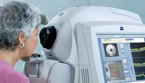
Optical coherence tomography (OCT) is one of the most common tests performed by a retinal specialist. OCT works essentially like an ultrasound, but instead of using sound waves, OCT uses light waves to obtain images of the retinal layers with higher resolution and magnification.
OCT scans the central retina (known as the macula) and provides a magnified cross-section view of the full thickness of the macula, much like looking at the slice of cake and all its layers from the side. These images of your macula reveal the presence of conditions such as macular edema, macular pucker, and macular holes, to name a few.
Repeat OCT scans are frequently used to follow retinal conditions and their response to treatment over time. OCT technology allows us to detect tiny amounts of fluid leakage and other abnormalities in the macula that are too small to be seen through direct visual examination alone.
What to expect during Optical Coherence Tomography testing
OCT is quick, painless, and non-invasive. This is not an X-Ray. Actually, the OCT machine is more like a sophisticated camera, and exposure to its light is not harmful to the eye. After you’re seated comfortably, you’re asked to place your chin in a chin-rest and then images are obtained.
The entire process takes just a few minutes. Once done, your doctor will review the images with you on a screen in your examination room.
Schedule your consultation with the internationally recognized retina specialists at Assil Gaur Eye Institute
The Spectralis® High-resolution OCT
AGEI is proud to offer one of the most advanced high-definition retinal imaging testing devices available today. The Heidelberg Scanning Laser OCT provides highly detailed, high-definition maps and images of the microscopic anatomy of the central retina (or macula) and other retinal structures, allowing us to make the most accurate diagnosis possible.
With automatic eye tracking and noise-reduction technology, the Spectralis® scanner has the ability to obtain the highest image resolution possible: right down to a few thousandths of a millimeter! What’s more, the Spectralis® performs the scan in a fraction of the time needed by other OCT scanners, making the process way more comfortable for our patients.
Ocular Ultrasound
Ocular ultrasound technology is used for imaging the anatomy of the eye when examining the inside of the eye by looking through the pupil is not possible. Such is the case in patients with a complete cataract, severe eye bleeding, or trauma. Ocular ultrasounds are also used to evaluate eye tumors.
What to expect during Ocular Ultrasound testing
Ocular ultrasonography is quick, painless, and non-invasive. Actually, it’s quite similar to ultrasounds performed on other parts of the body. You’re seated comfortably and asked to close your eyes because your eyelids do not have to be open for this test. Your doctor then uses a wand to gently swab cold, sterile gel solution over your eyelid as the scanner captures images of the eye.
The entire process takes only a few minutes. Your doctor will review the results with you on the screen in your examination room immediately following the test.
Experience Assil Gaur Eye Institute's world-class retina care
The AGEI staff includes a highly-skilled retina specialist Dr. Svetlana Pilyugina or “Dr. P”, as she is known to her patients. Dr. P is a Stanford educated and fellowship-trained ophthalmologist. She is board certified in diseases and surgery of the vitreous and retina.
Dr. Pilyugina has extensive experience in the treatment of floaters, flashes, and all retinal conditions. To schedule an appointment, either call 866-945-2745 or click here to make an appointment online.


