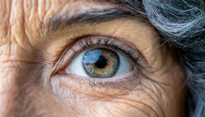
What is Macular Degeneration (AMD)?
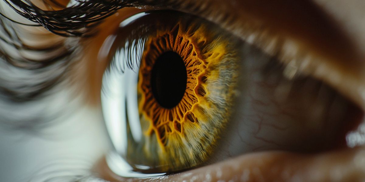
Macular Degeneration is an eye condition that occurs when aging causes damage to the macula. The macula is part of the retina (the light-sensitive tissue at the back of the eye that controls sharp, straight-ahead vision.
Age-related macular degeneration (AMD) is the leading cause of irreversible blindness in adults over 55. It is expected to affect 288 million people worldwide by the year 2040.
- What causes Macular Degeneration?
- What are the symptoms of macular degeneration?
- Is there a cure for macular degeneration?
- There are two types of macular degeneration. How are they different?
- Can you prevent macular degeneration?
- Experience Assil Gaur Eye Institute's nationally recognized macular degeneration care.
- MACULAR DEGENERATION FAQs
- Is macular degeneration hereditary?
- What are the early warning signs of macular degeneration?
- Can macular degeneration be reversed?
- What does vision look like with macular degeneration?
- What is the best eye vitamin for macular degeneration?
- Which is more severe wet or dry macular degeneration?
- How fast does macular degeneration progress?
- How is macular degeneration diagnosed?
- What foods should be avoided with macular degeneration?
- What percentage of macular degeneration patients go blind?
- Are there two forms of wet AMD?
- Can you have both glaucoma and macular degeneration?
What causes Macular Degeneration?

The exact cause of macular degeneration is not exactly known, but research indicated it’s a combination of genetic and environmental factors. These risk factors include:
- Having a family history of AMD
- Age (most cases are diagnosed after age 55)
- Smoking (Studies show that smoking is associated with 25% of severe AMD cases, and living with a smoker doubled test subjects' risk of developing AMD)
- Obesity
- Being Caucasian
- Being female
- If you take certain medications like chloroquine or phenothiazine-type drugs.
What are the symptoms of macular degeneration?
- Loss of central vision or dark spots in the center of vision
- A well-defined blind spot in your field of vision
- Distorted vision, with straight lines appearing wavy
- Decreased brightness of colors and overall haziness in your vision
- Difficulty adjusting to changes in lighting
- Needing more light to perform tasks
- Loss of depth perception
- Blurred vision
Is there a cure for macular degeneration?
No. However, the last decade has made significant breakthroughs in treatment options. Today, some medications help control and slow the progression of AMD and, in some cases, may even improve vision.
There are two types of macular degeneration. How are they different?
The two forms of macular degeneration are called dry and wet macular degeneration. Both affect the macula, which is part of the retina of your eye. Their differences lie in the speed of the onset of the disease and its severity.
What is dry macular degeneration?
Dry AMD is the less severe early stage and is more common. Eighty percent of AMD patients have a dry form, which gradually worsens over the years.
In the dry stage, tiny yellow deposits of metabolic waste, known as ¨drusen¨, form in the macula. At first, they have little effect on vision. However, as the disease progresses and drusen accumulate, they obstruct the flow of nutrients and oxygen to the macula.
How is dry macular degeneration treated?
The only treatment available for dry AMD is a pill called AREDS2 formula. AREDS2 is a mixture of antioxidant supplements and two visual pigments (lutein and zeaxanthin) essential for retinal function.
AREDS2 has been shown to reduce the risk of dry AMD progressing to the advanced Wet form by 25% per year.
What is wet macular degeneration?
Wet macular degeneration (Wet AMD) is the more advanced form of this disease, and about 20% of Dry AMD patients will progress to it. Also, If the condition has quickly appeared over weeks to months, it too is wet AMD.
The wet stage of AMD is aggressive and requires urgent treatment by a retinal specialist to prevent progressive vision loss. If wet AMD is left untreated, blood or liquid build-up in the retina leads to scarring, which can cause irreversible vision loss.
Eyes with wet AMD have increased production of a Vascular Endothelial Growth Factor (VEGF) protein. This protein triggers the growth of new blood vessels in the retina. Too much VEGF in the eye leads to the creation of fragile malformed blood vessels. These vessels leak, causing damage to the macula and surrounding retina that eventually leads to central vision loss.
If you get wet AMD in one eye, you’ll have a 20% chance of developing it in your other eye within five years.
How is Wet Macular Degeneration treated?
Once AMD progresses to the wet form, pills are no longer an acceptable treatment option. Instead, medications must be placed directly within the eye to help control the disease at its source. Luckily, injectable medications have revolutionized the treatment for Wet AMD.
Although they sound scary, intraocular injections are quick, painless, safe, and effective in slowing disease progression. And in some cases, restoring some of the lost vision, Known as anti-VEGF medications, which include Lucentis and Eyelea.
In some cases, a laser treatment called photodynamic therapy may be effective. Our retina specialist will recommend the option(s) best suited for your situation.
Can you prevent macular degeneration?
No, but simple lifestyle changes can decrease your risk of developing it. These include eating plenty of dark green leafy vegetables, salmon, and other cold-water fish (for their omega-3 fatty acids).
It's also essential to exercise and avoid high blood pressure and high cholesterol. Wear sunglasses that provide 100 percent UV protection and protect your eyes from visible radiation and blue light. Lastly, if you smoke, stop!
We can’t overstate the importance of getting an annual eye exam to screen for AMD and other eye health conditions that can threaten your vision and quality of life.
Schedule your consultation today with our internationally recognized retina specialists
Experience Assil Gaur Eye Institute's nationally recognized macular degeneration care.
The AGEI staff includes a highly skilled Stanford-trained retina specialist, Dr. Svetlana Pilyugina (or “Dr. P”, as she is known to her patients). Dr. P is an ophthalmologist with fellowship training and board certification in diseases and surgery of the vitreous and retina.
Dr. Pilyugina has extensive experience treating macular degeneration, retinal detachment, and diabetic retinopathy, having treated thousands of patients for over a decade.
As a member of an elite group of only 3000 vitreoretinal specialists in the United States, Dr. Pilyugina brings to AGEI a unique skill set in the treatment and surgery of retinal disease. Her academic credentials include numerous research papers, conference presentations, medical publications, and ophthalmology clinical trials.
We are conveniently located for patients throughout Southern California and the Los Angeles area in or near Beverly Hills, Santa Monica, West Los Angeles, West Hollywood, Culver City, Hollywood, Venice, Marina del Rey, Malibu, Manhattan Beach, and Downtown Los Angeles.
MACULAR DEGENERATION FAQs
Is macular degeneration hereditary?
Yes, macular degeneration can have a hereditary component. However, having a genetic predisposition to the disease does not necessarily mean an individual will develop macular degeneration. Lifestyle factors such as smoking, diet, and sun exposure can also increase the risk of developing the disease.
What are the early warning signs of macular degeneration?
The early warning signs of macular degeneration may include:
- Objects appear distorted or blurry, or that straight lines look wavy.
- Need help seeing in dimly lit environments, such as restaurants or movie theaters.
- Colors appear less vivid, or it is difficult to distinguish between shades of colors, and there is an inability to see fine details.
- A dark spot in the center of your visual field.
- Prolonged adaptation to changes in lighting.
Can macular degeneration be reversed?
No, it cannot be reversed. However, early detection and treatments can slow or pause the progression of the disease, allowing you to maintain your lifestyle during treatments.
What does vision look like with macular degeneration?
Macular degeneration can cause blurry or distorted vision in the center of the visual field. Usually, you're still able to use your peripheral vision. You can learn how to use your side vision to help you do things with the help of a vision rehabilitation specialist who can teach you how this works.
What is the best eye vitamin for macular degeneration?
No specific vitamin or nutritional supplement has been proven to cure or prevent macular degeneration. However, a large study by the National Eye Institute, called AREDS (Age-Related Eye Disease Study), found that certain vitamins and nutrients like beta-carotene and vitamin E may protect the eyes in some people.
Which is more severe wet or dry macular degeneration?
Wet (exudative) macular degeneration is generally considered more severe and can progress faster than dry (atrophic)macular degeneration.
How fast does macular degeneration progress?
In general, dry macular degeneration can take years or even decades to progress to the more advanced stages, where central vision loss becomes more noticeable.
Wet macular degeneration can progress rapidly and cause severe vision loss in weeks or months if left untreated.
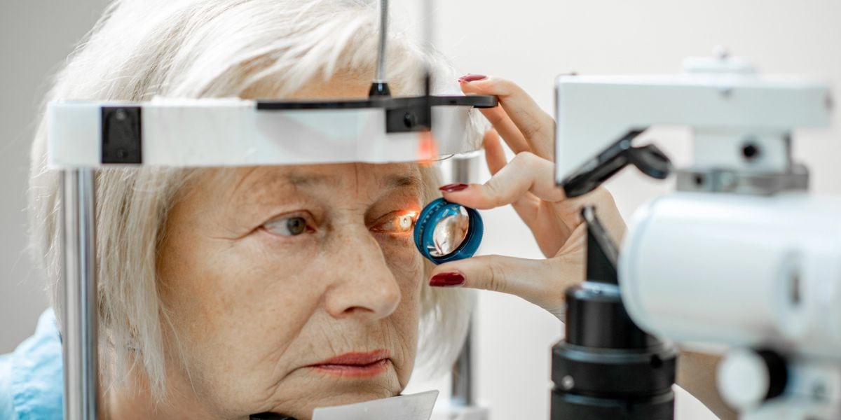
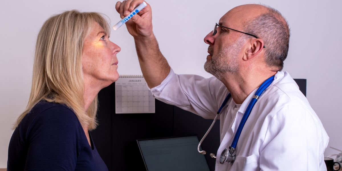
How is macular degeneration diagnosed?
Macular degeneration is typically diagnosed through a comprehensive eye exam that may include the following tests:
- Visual acuity test: Using an eye chart, this test measures how well you can see at different distances.
- Dilated eye exam: During this exam, your eye care provider will use dilating eye drops to widen your pupils and examine the back of your eye, including the macula, for signs of macular degeneration and other eye conditions.
- An Amsler grid test allows the patient to look at a grid, identify all four corners, and the doctor to analyze the response for distortion or missing areas in the central vision.
- Optical coherence tomography (OCT): This non-invasive imaging test uses light waves to create a detailed cross-sectional image of the retina, allowing your eye doctor to see any changes or damage to the macula.
- Fluorescein angiography: In this test, a dye is injected into a vein in your arm. Your eye doctor will take photographs of your retina as the dye circulates through your blood vessels. This can help identify abnormal blood vessels or leakage causing wet macular degeneration.
What foods should be avoided with macular degeneration?
Foods that should be avoided or limited include:
- Processed and fried foods: These foods are high in unhealthy fats, contributing to inflammation and increasing the risk of macular degeneration.
- Sugary and high-glycemic-index foods: These foods can cause spikes in blood sugar levels, contributing to inflammation and increasing the risk of macular degeneration.
- Red and processed meats: These foods are high in saturated fats and can contribute to inflammation and increase the risk of macular degeneration.
- Trans fats: These are found in many processed and baked goods, contributing to inflammation and increasing the risk of macular degeneration.
What percentage of macular degeneration patients go blind?
Most people with macular degeneration do not go completely blind. Still, they may experience significant low vision, making it difficult or virtually impossible to perform everyday activities like reading, driving, and recognizing faces.
Are there two forms of wet AMD?
Yes, there are two forms of Wet AMD: occult and classic.
Occult wet macular degeneration is when new blood vessels grow less, and their leakage is less evident and is associated with less severe vision loss. Classic wet macular degeneration is when blood vessels and scars are clearly visible on the retina and usually results in more severe vision loss.
Can you have both glaucoma and macular degeneration?
Yes, a person can have both glaucoma and macular degeneration. Although these are two different eye conditions, they can coincide, especially in older adults.
Research Sources:
AREDS 2 Supplements for Age-Related Macular Degeneration (AMD) | National Eye Institute | https://www.nei.nih.gov/learn-about-eye-health/eye-conditions-and-diseases/age-related-macular-degeneration/nutritional-supplements-age-related-macular-degeneration
Age-Related Macular Degeneration (AMD) | National Eye Institute | https://www.nei.nih.gov/learn-about-eye-health/eye-conditions-and-diseases/age-related-macular-degeneration
Vision Loss, Central - American Academy of Ophthalmology | https://www.aao.org/eye-health/symptoms/vision-loss-central
Ocular nutritional supplements: are their ingredients and manufacturers' claims evidence-based? PubMed | https://pubmed.ncbi.nlm.nih.gov/25458196
Dietary fatty acid intake, plasma fatty acid levels, and the risk of age-related macular degeneration (AMD): a dose-response meta-analysis of prospective cohort studies - PubMed | https://pubmed.ncbi.nlm.nih.gov/33469697/
Macular degeneration: peculiar sunlight exposure in an agricultural worker - PMC
https://www.ncbi.nlm.nih.gov/pmc/articles/PMC7812542/
Omega 3 fatty acids for preventing or slowing the progression of age‐related macular degeneration | https://www.ncbi.nlm.nih.gov/pmc/articles/PMC7087473/
Articles from our blog on macular degeneration
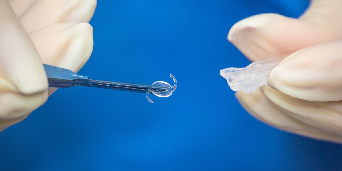
- Breakthrough Eye Implant Technology Offers New Hope for Patients with Advanced Macular Degeneration
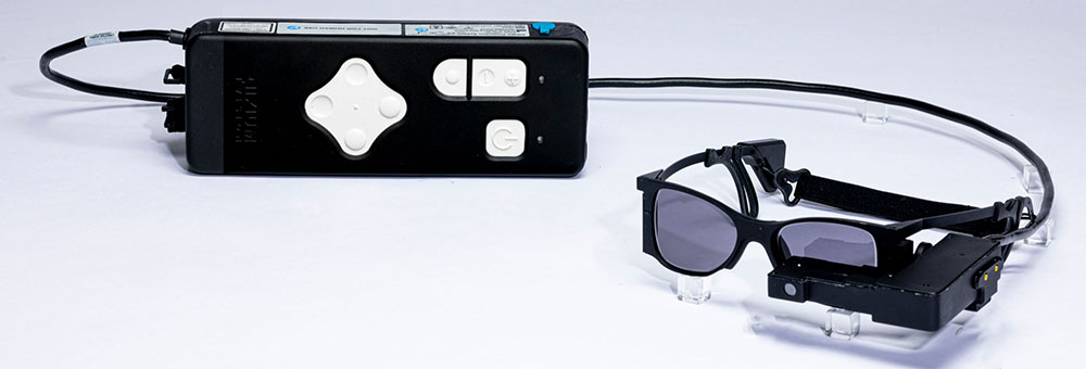
- Valeda Light Delivery System for Dry Age-Related Macular Degeneration (AMD)
- Does High Blood Pressure Affect Your Vision?
- 7 Ways to Adapt Your Home for Vision Loss
- Could Age-Related Vision Loss Be Reversed? New Tests Look Promising
- The Gender Health Gap: Do Women Have Worse Vision?
- What's the Worst Food for your Eyes? Beware These 5
- Prozac Is a Potential Treatment for Age-Related Macular Degeneration [new study]
- LED Lights Can Harm Your Eyes
- Can Green Smoothies Support Eye Health?
-
Retina Specialist
As a member of an elite group of only 3000 retina-vitreous specialists in the United States, Stanford trained Dr. Pilyugina brings to AGEI a unique skill set in the treatment and surgery of retinal disease. Her academic credentials include numerous research papers, conference presentations, medical publications, and clinical trials.

 Excellent experience. Kerry Assil is on the cutting edge of eye technology. He has surrounded himself with very capable and pat...Brent T.
 Their whole staff is fabulous and so caring!!! The Annual Eye Exam that Dr May Performs is so extensive and done with such...Joan M.
- I am pleased with the service I have received. And I will continue to recommend to my friends and familyFelix C.
- The staff is personable and professional.Kurt E.
- Great service, everyone was super friendly. I was originally really nervous about getting Lasik but both Dr. Assil, Dr. Natalie...Peter S.
- Dr may is greatJennifer L.
- Very good treatment!Steven M.
- NopeCaroline M.
- Great care, as normal.Ken H.
- Always too long of a waitCindra L.
- MD was very attentive to my concerns.Charles D.
- Everything was great, not much else I can sayBarbara M.
-
 Listen Now
Can Vitamin D Help Relieve Dry Eye Disease? A New Study Says Yes!
Read
Listen Now
Can Vitamin D Help Relieve Dry Eye Disease? A New Study Says Yes!
Read
-
 Listen Now
What Are the Causes of Double Vision and How Can They Be Addressed?
Read
Listen Now
What Are the Causes of Double Vision and How Can They Be Addressed?
Read
-
 Listen Now
Patients are raving about the enVista® Envy™ Trifocal IOL Lens
Read
Listen Now
Patients are raving about the enVista® Envy™ Trifocal IOL Lens
Read
-
 Listen Now
Are Cataracts at a Young Age Getting More Common?
Read
Listen Now
Are Cataracts at a Young Age Getting More Common?
Read
-
 Listen Now
Can Stress Increase Eye Pressure in Glaucoma Patients?
Read
Listen Now
Can Stress Increase Eye Pressure in Glaucoma Patients?
Read
-
 Kerry and Tanaz Assil focus on the fruits of their labor
Read
Kerry and Tanaz Assil focus on the fruits of their labor
Read
-
 Breakthrough Eye Implant Technology Offers New Hope for Patients with Advanced Macular Degeneration
Read
Breakthrough Eye Implant Technology Offers New Hope for Patients with Advanced Macular Degeneration
Read
-
 Listen Now
Cataract Lens Replacement Failure: What Can You Do About It?
Read
Listen Now
Cataract Lens Replacement Failure: What Can You Do About It?
Read
-
 Listen Now
What are Some Common Medications that can Cause or Worsen Glaucoma?
Read
Listen Now
What are Some Common Medications that can Cause or Worsen Glaucoma?
Read
-
 Listen Now
Understanding Pinguecula: Causes, Symptoms, and Treatment
Read
Listen Now
Understanding Pinguecula: Causes, Symptoms, and Treatment
Read
-
 How Autoimmune Diseases Affect Your Eyes: What You Need to Know
Read
How Autoimmune Diseases Affect Your Eyes: What You Need to Know
Read
-
 Listen Now
Can Pupil Size Reveal Intelligence? Maybe Size Really Does Matter!
Read
Listen Now
Can Pupil Size Reveal Intelligence? Maybe Size Really Does Matter!
Read
-
 Valeda Light Delivery System for Dry Age-Related Macular Degeneration (AMD)
Read
Valeda Light Delivery System for Dry Age-Related Macular Degeneration (AMD)
Read
-
 Listen Now
Alcohol and Dry Eye Syndrome: Understanding the Connection
Read
Listen Now
Alcohol and Dry Eye Syndrome: Understanding the Connection
Read
-
 Listen Now
The History of IOLs: You’ve Come A Long Way, Baby!
Read
Listen Now
The History of IOLs: You’ve Come A Long Way, Baby!
Read


Look Who Trusts Their Eyes to Assil Gaur Eye Institute...
-
LeBron James
Los Angeles LakersLeBron James is one of the NBA's greatest of all time. He absolutely depends on his vision to perform at the very highest level. That's why he's an AGEI LASIK surgery patient.
-
Anthony Davis
Dallas Mavericks -
Chris Paul
San Antonio Spurs -
Paul George
Philadelphia 76ers -
Marlee Matlin
Actress -
Michelle Williams
Actress -
Dwyane Wade
Miami Heat (retired) -
LaToya Jackson
Entertainer -
Lorenzo Lamas
Actor -
Philip Bailey
Earth Wind and Fire -
Gary Sinise
Actor -
Rip Hamilton
Former NBA player -
John Salley
Commentator, former NBA player -
Blair Underwood
Actor -
Tom ArnoldComedian/Actor
-
Troy EvansActor
-
John NobleActor
-
Isaac EddyActor/Director/Teacher
-
Barbara RushActress
-
John EmersonUS Ambassador to Germany
-
Shane MosleyFormer professional boxer
-
Cuttino MobleyFormer NBA player
-
Seth GordonDirector
-
Maurice EvansFormer NBA player
-
Wang LuoyongActor
-
Jesse MetcalfeActor
-
Ananda LewisTelevision Personality
-
Rob BrydonComedian
-
Clifton Collins Jr.Actor
-
Malinda WilliamsActress
-
Adrienne FrantzActress
-
Brad PittActor
-
LeBron JamesLos Angeles Lakers
-
Anthony DavisDallas Mavericks
-
Chris PaulSan Antonio Spurs
-
Courtney CoxActress
-
Paul GeorgePhiladelphia 76ers
-
Marlee MatlinActress
-
Michelle WilliamsActress
-
Dwyane WadeMiami Heat (retired)
-
Jalen BrunsonNew York Knicks
-
Mychal ThompsonCommentator, former NBA player
-
LaToya JacksonEntertainer
-
Lorenzo LamasActor
-
Philip BaileyEarth Wind and Fire
-
Gary SiniseActor
-
Rip HamiltonFormer NBA player
-
John SalleyCommentator, former NBA player
-
Blair UnderwoodActor
-
Tom ArnoldComedian/Actor
-
Troy EvansActor
-
John NobleActor
-
Isaac EddyActor/Director/Teacher
-
Barbara RushActress
-
Rudy GarciduenasBlue Man Group
-
John EmersonUS Ambassador to Germany
-
Shane MosleyFormer professional boxer
-
Cuttino MobleyFormer NBA player
-
Seth GordonDirector
-
Maurice EvansFormer NBA player
-
Cheri BlauwetPhysician and Wheelchair Racer
-
Wang LuoyongActor
-
Jesse MetcalfeActor
-
Jeana WilsonActress
-
Courtnee DraperActress
-
Ananda LewisTelevision Personality
-
Robyn JohnsonProducer
-
Rob BrydonComedian
-
Cary SchumanActor
-
Roland Buddy Lewis Jr.Comedian
-
Clifton Collins Jr.Actor
-
Malinda WilliamsActress
-
Chad JeffersMusician
-
Adrienne FrantzActress
-
Franco CarlottoBody Builder
-
Brad MatesMusician
-
David PichetteMusician
-
Mike MelaconProfessional Baseball Player
-
Nautica de la CruzRadio Personality
-
Lynn ConkwrightBody Builder
-
Nely GalanProducer
-
Roxanne GallaActress