
What is Pterygium (surfer’s eye)?
A pterygium is a raised fleshy growth filled with blood vessels that originate in the conjunctiva (the clear membrane covering the white of the eye) and spreads over the cornea (the clear outer covering of your eye).
A pterygium can range from a transparent area with a few blood vessels to a thick, opaque growth that can obstruct vision. It can occur in one or both eyes and typically originates in the inner corner of the eye and spreads toward the pupil.
- What are Pterygium Symptoms?
- Pterygium causes and risk factors
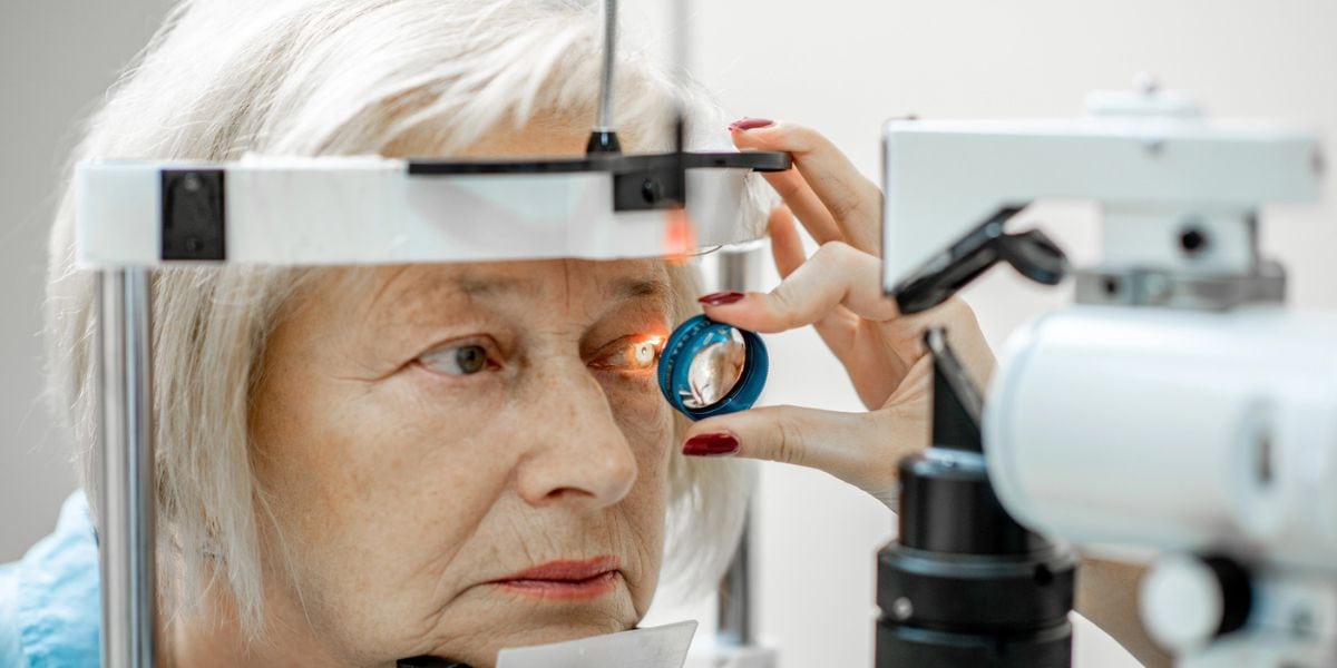
- How is pterygium diagnosed?
- Pterygium treatment
- Pterygium surgery (pterygium excision)
- How does AGEI cut pterygium recurrence from 80% to below 1%?
- What to expect after eye whitening surgery
- Should I undergo pterygium removal eye surgery?
- Assil Gaur Eye Institute, the best choice for pterygium surgery near you
- Pterygium FAQs
- Can surfer’s eye go away?
- How to prevent surfer’s eye (pterygium)?
- Is pterygium dangerous?
- Why is it called surfer’s eye?
- Is pterygium sometimes confused with pinguecula?
- What are the risks of a pterygium surgical procedure?
What are Pterygium Symptoms?
Pterygium is usually asymptomatic in its early stages. However, when inflamed, it can cause eye irritation: itching, tearing, and burning. As the pterygium advances, it can grow over your iris and pupil, causing blurred vision. A pterygium can also cause contact lens intolerance.
Pterygium causes and risk factors
pterygium, its causes, symptoms, and treatments.
The exact cause of pterygium is unclear, but it is found more often in populations living close to the equator who have prolonged exposure to sun and UV rays. This condition is sometimes called Surfer’s Eye because of its common occurrence among surfers.
Risk factors for pterygium include:
- Exposure to ultraviolet light (from the sun or other sources)
- Residing in sunny climates close to the equator
- Being in dry, dusty climates
- Spending a lot of time outdoors (working outdoors increases your risk by 150%)
- Complications from dry eye disease
How is pterygium diagnosed?
By simple observation. We can make a formal diagnosis following a slit-lamp examination that allows close-up observation of the lesion under magnification. A biopsy is often taken at the time of removal and sent to a pathologist for diagnostic confirmation.
If you have a pterygium, a thorough eye exam should be performed to assess its impact on your vision and rule out less common diagnoses that can cause an eye tumor.
Our eye specialist will also measure how far the pterygium extends over your cornea. Usually, you will return for follow-up exams every 1 to 2 years to determine the rate of its growth toward your visual axis.
Pterygium treatment
Treatment may not be required if your symptoms aren’t causing discomfort or interfering with your vision.
Our eye care provider may:
- Recommend over-the-counter anti-allergy drops, lubricating eye drops, artificial tears, anti-inflammatory agents, and ointments.
- Prescribe steroid drops or ointments to reduce pain, redness, itching, and swelling.
- Recommend wearing UVA/UVB-blocking sunglasses when outdoors.
Schedule your consultation today with our internationally recognized eye surgeons
Pterygium surgery (pterygium excision)
If conservative treatments aren’t successful in relieving symptoms, or the appearance of pterygia makes you self-conscious, surgery may be needed.
Unfortunately, pterygium surgery is not as simple as cutting the growth out of your conjunctiva because this approach is associated with an 80 percent chance of the pterygium growing back. In fact, 97 percent of regrowth occurs in the first year following the surgical removal of a pterygium.
Because of the high recurrence rate associated with simple pterygium extraction, our ophthalmologists sometimes remove some of the patient's conjunctiva from another part of the eye and use it as a graft to fill in the gap left when the pterygium is removed.
This approach, known as conjunctival autograft, continues to be used with success. However, recurrence rates can still be up to 33 percent. Although the odds are better, they are still not great.
How does AGEI cut pterygium recurrence from 80% to below 1%?
Over the past 12 years, AGEI's surgeons have adopted many advancements in the field, which has led to a pterygium recurrence rate below 1 percent in our practice. These surgical techniques include:
- Carefully removing the pterygium and underlying scar tissue to achieve clean borders
- Minimizing the surgical manipulation of the tissue surrounding the gap
- Using amniotic membrane graft for its ability to suppress post-op inflammation, scarring and new blood vessel formation
- Bathing the gap created by the pterygium removal with two powerful medications to minimize post-op inflammation and scarring that lead to increased risk of recurrence
- Affixing the graft with fibrin glue to decrease inflammation and irritation at the graft site caused by sutures and to act as a physical barrier along the perimeter of the graft site to thwart pterygium re-growth
- Following a meticulous post-op medication regimen designed to suppress swelling and infection
What to expect after eye whitening surgery
After surgery, your doctor will prescribe steroids and antibiotic eye drops to minimize the chances of recurrence and prevent infection. Compliance with the dosing schedule is key to helping prevent recurrent pterygium.
Your doctor will check you on the day following surgery. Then you will follow up at one week, one month, three months, six months, and one year post-op. After that, you will be seen annually to check for pterygium recurrence.
You may feel irritation and a foreign body sensation in your eye once your anesthesia wears off. These symptoms will resolve in a week. There will also be redness at the surgical site that can last up to six weeks.
Typically, patients take six to eight weeks to heal fully. Smaller pterygia heal faster than larger ones.
Should I undergo pterygium removal eye surgery?
With the dramatic improvement in results over the years at our center, most patients now undergo removal at earlier stages for cosmetic reasons rather than due to any visual loss.
Still, the decision to undergo pterygium removal surgery should be made after careful consideration and discussion with your eye doctor and primary health care provider to ensure that you understand the risks and benefits of the procedure and are willing to perform the post-operative eye care regimen necessary for successful results.
Assil Gaur Eye Institute, the best choice for pterygium surgery near you
Our nationally recognized ophthalmologists and corneal specialists are leaders in a wide range of ophthalmological conditions, including state-of-the-art LASIK vision correction, retinal treatments, cataract surgery, glaucoma care, macular disease, and diabetic eye conditions, to name just a few.
We are conveniently located for patients throughout Southern California and the Los Angeles area at locations in or near Los Angeles, Beverly Hills, Santa Monica, West Los Angeles, West Hollywood, Culver City, Hollywood, Venice, Marina del Rey, Malibu, Manhattan Beach, and Downtown Los Angeles.
![]()
Pterygium FAQs
Can surfer’s eye go away?
Surfer's eye can go away with treatment or surgery, but preventive measures are crucial to avoid recurrence.
How to prevent surfer’s eye (pterygium)?
To prevent surfer's eye, you can take the following steps:
- Wear sunglasses that block 100% of UV rays to protect your eyes from the harmful effects of the sun. Look for sunglasses with polarized lenses that reduce glare and provide better vision on the water.
- Wear a wide-brimmed hat to shade your face and eyes from the sun's rays.
- Use artificial tears to keep your eyes moist and prevent dryness, which can lead to irritation.
- Take regular breaks from being in the sun, wind, and dust to give your eyes a rest.
- Rubbing your eyes can irritate them and increase the risk of developing surfer's eye.
- Smoking can increase the risk of developing surfer's eye and other eye diseases.
Is pterygium dangerous?
Because it is referred to as a tumor, some people may fear it is a form of cancer. Rest assured, pterygium is a benign (non-cancerous growth) lesion that does not spread beyond the eye's surface. In some cases, it can also lead to astigmatism or distortion of the cornea, which can affect the clarity of vision.
Why is it called surfer’s eye?
Surfers' eyes, also known as pterygium, are called that because they are often seen in people who spend a lot of time in the sun and wind, such as surfers. The condition is most commonly seen in people who live in tropical or subtropical climates with high ultraviolet radiation exposure.
Is pterygium sometimes confused with pinguecula?
Yes. Both pinguecula and pterygium are conditions that affect the conjunctiva, the clear tissue covering the white part of the eye and the inside of the eyelids. However, they are different in their characteristics and symptoms.
A pinguecula is a yellowish patch or bump on the conjunctiva, often on the side of the eye nearest the nose. It is caused by a change in normal tissue that deposits protein, fat, or calcium. It's similar to a callus on the skin. Pingueculas are often seen in middle-aged or older people who have had extensive exposure to sunlight. However, they can also appear in younger people and children, particularly those who spend a lot of time under the sun without proper eye protection.
Pinguecula often don't cause symptoms, but they can cause discomfort, a feeling of something in the eye, or they can become inflamed and cause dry eye symptoms.
What are the risks of a pterygium surgical procedure?
Pterygium surgery generally has good outcomes, particularly with patients who comply with their post-op prescription eye drop schedule. There are some rare risks associated with pterygium surgery. These include:
- Eye swelling
- Double vision
- Prolonged redness
- Infection
Sources
Necrotising scleritis after bare sclera excision of pterygium. Alsagoff Z, Tan DTH, Chee S. Br J Ophthalmol. (https://www.ncbi.nlm.nih.gov/pmc/articles/PMC1723636/) 2000;84:1050
What is a Pinguecula and a Pterygium (Surfer’s Eye)? Academy of Ophthalmology. EyeSmart. (https://www.aao.org/eye-health/diseases/pinguecula-pterygium) Accessed 2/22/2022
Consumer Version. Pinguecula and Pterygium. (https://www.merckmanuals.com/home/eye-disorders/conjunctival-and-scleral-disorders/pinguecula-and-pterygium?query=pterygium)
-

Dr. May is an optometrist who has worked with Dr. Assil and the Assil Gaur Eye Institute for over a dozen years . He provides a broad range of care for patients including pre and post-surgical management, comprehensive eye exams, urgent care as well as dry eye consultation and therapy. He's also assisted in numerous FDA supervised clinical trials run at AGEI.

 Great experience. Amazing results. Dr. Adele stays connected through entire experience. Fully recommend.Hans C.
- Dr. Assil is the best eye doctor ever. My daughter had an eye issue and his staff went out of their way to get her seen and we...K B.
- This by far the most Professional Organization for eyes probably in the World. They are Caring, Thoughtful, do Amazing work and...Philip B.
 Their whole staff is fabulous and so caring!!! The Annual Eye Exam that Dr May Performs is so extensive and done with such...Joan M.
- I am pleased with the service I have received. And I will continue to recommend to my friends and familyFelix C.
- The staff is personable and professional.Kurt E.
- Dr may is greatJennifer L.
- Very good treatment!Steven M.
- NopeCaroline M.
- Always too long of a waitCindra L.
- MD was very attentive to my concerns.Charles D.
- Everything was great, not much else I can sayBarbara M.
-
 Listen Now
Can Pupil Size Reveal Intelligence? Maybe Size Really Does Matter!
Read
Listen Now
Can Pupil Size Reveal Intelligence? Maybe Size Really Does Matter!
Read
-
 Listen Now
Understanding Pinguecula: Causes, Symptoms, and Treatment
Read
Listen Now
Understanding Pinguecula: Causes, Symptoms, and Treatment
Read
-
 Kerry and Tanaz Assil focus on the fruits of their labor
Read
Kerry and Tanaz Assil focus on the fruits of their labor
Read
-
 Listen Now
Can Stress Increase Eye Pressure in Glaucoma Patients?
Read
Listen Now
Can Stress Increase Eye Pressure in Glaucoma Patients?
Read
-
 Listen Now
What Are the Causes of Double Vision and How Can They Be Addressed?
Read
Listen Now
What Are the Causes of Double Vision and How Can They Be Addressed?
Read
-
 Listen Now
Cataract Lens Replacement Failure: What Can You Do About It?
Read
Listen Now
Cataract Lens Replacement Failure: What Can You Do About It?
Read
-
 Listen Now
Alcohol and Dry Eye Syndrome: Understanding the Connection
Read
Listen Now
Alcohol and Dry Eye Syndrome: Understanding the Connection
Read
-
 Valeda Light Delivery System for Dry Age-Related Macular Degeneration (AMD)
Read
Valeda Light Delivery System for Dry Age-Related Macular Degeneration (AMD)
Read
-
 Listen Now
Are Cataracts at a Young Age Getting More Common?
Read
Listen Now
Are Cataracts at a Young Age Getting More Common?
Read
-
 Listen Now
What are Some Common Medications that can Cause or Worsen Glaucoma?
Read
Listen Now
What are Some Common Medications that can Cause or Worsen Glaucoma?
Read
-
 Breakthrough Eye Implant Technology Offers New Hope for Patients with Advanced Macular Degeneration
Read
Breakthrough Eye Implant Technology Offers New Hope for Patients with Advanced Macular Degeneration
Read
-
 Listen Now
Can Vitamin D Help Relieve Dry Eye Disease? A New Study Says Yes!
Read
Listen Now
Can Vitamin D Help Relieve Dry Eye Disease? A New Study Says Yes!
Read
-
 Listen Now
The History of IOLs: You’ve Come A Long Way, Baby!
Read
Listen Now
The History of IOLs: You’ve Come A Long Way, Baby!
Read
-
 Listen Now
Patients are raving about the enVista® Envy™ Trifocal IOL Lens
Read
Listen Now
Patients are raving about the enVista® Envy™ Trifocal IOL Lens
Read
-
 How Autoimmune Diseases Affect Your Eyes: What You Need to Know
Read
How Autoimmune Diseases Affect Your Eyes: What You Need to Know
Read


Look Who Trusts Their Eyes to Assil Gaur Eye Institute...
-
LeBron James
Los Angeles LakersLeBron James is one of the NBA's greatest of all time. He absolutely depends on his vision to perform at the very highest level. That's why he's an AGEI LASIK surgery patient.
-
Anthony Davis
Dallas Mavericks -
Chris Paul
San Antonio Spurs -
Paul George
Philadelphia 76ers -
Marlee Matlin
Actress -
Michelle Williams
Actress -
Dwyane Wade
Miami Heat (retired) -
LaToya Jackson
Entertainer -
Lorenzo Lamas
Actor -
Philip Bailey
Earth Wind and Fire -
Gary Sinise
Actor -
Rip Hamilton
Former NBA player -
John Salley
Commentator, former NBA player -
Blair Underwood
Actor -
Tom ArnoldComedian/Actor
-
Troy EvansActor
-
John NobleActor
-
Isaac EddyActor/Director/Teacher
-
Barbara RushActress
-
John EmersonUS Ambassador to Germany
-
Shane MosleyFormer professional boxer
-
Cuttino MobleyFormer NBA player
-
Seth GordonDirector
-
Maurice EvansFormer NBA player
-
Wang LuoyongActor
-
Jesse MetcalfeActor
-
Ananda LewisTelevision Personality
-
Rob BrydonComedian
-
Clifton Collins Jr.Actor
-
Malinda WilliamsActress
-
Adrienne FrantzActress
-
Brad PittActor
-
LeBron JamesLos Angeles Lakers
-
Anthony DavisDallas Mavericks
-
Chris PaulSan Antonio Spurs
-
Courtney CoxActress
-
Paul GeorgePhiladelphia 76ers
-
Marlee MatlinActress
-
Michelle WilliamsActress
-
Dwyane WadeMiami Heat (retired)
-
Jalen BrunsonNew York Knicks
-
Mychal ThompsonCommentator, former NBA player
-
LaToya JacksonEntertainer
-
Lorenzo LamasActor
-
Philip BaileyEarth Wind and Fire
-
Gary SiniseActor
-
Rip HamiltonFormer NBA player
-
John SalleyCommentator, former NBA player
-
Blair UnderwoodActor
-
Tom ArnoldComedian/Actor
-
Troy EvansActor
-
John NobleActor
-
Isaac EddyActor/Director/Teacher
-
Barbara RushActress
-
Rudy GarciduenasBlue Man Group
-
John EmersonUS Ambassador to Germany
-
Shane MosleyFormer professional boxer
-
Cuttino MobleyFormer NBA player
-
Seth GordonDirector
-
Maurice EvansFormer NBA player
-
Cheri BlauwetPhysician and Wheelchair Racer
-
Wang LuoyongActor
-
Jesse MetcalfeActor
-
Jeana WilsonActress
-
Courtnee DraperActress
-
Ananda LewisTelevision Personality
-
Robyn JohnsonProducer
-
Rob BrydonComedian
-
Cary SchumanActor
-
Roland Buddy Lewis Jr.Comedian
-
Clifton Collins Jr.Actor
-
Malinda WilliamsActress
-
Chad JeffersMusician
-
Adrienne FrantzActress
-
Franco CarlottoBody Builder
-
Brad MatesMusician
-
David PichetteMusician
-
Mike MelaconProfessional Baseball Player
-
Nautica de la CruzRadio Personality
-
Lynn ConkwrightBody Builder
-
Nely GalanProducer
-
Roxanne GallaActress