Keratoconus Progression: Stages, Acceleration, and Treatment Decisions
Keratoconus progression is most clearly seen through close-up corneal imaging, where the hallmark forward bulging of the cone-shaped cornea becomes unmistakable—a structural change that, left unmonitored, quietly steals clarity long before a patient notices dramatic vision loss.
Advanced tomographic maps produced at elite practices like Assil Gaur Eye Institute capture micron-level distortions in corneal curvature, giving our world-class specialists the precision data needed to intervene at exactly the right moment. Trusting that level of detail to anything less than a dedicated corneal expert is a risk no patient should take.
If your vision has been getting worse every year and your glasses prescription keeps changing, keratoconus progression may already be reshaping your cornea – and the window to stop it is narrower than most patients realize. That’s not meant to alarm you. It’s meant to create the urgency most general eye exams fail to communicate.
We understand the frustration: you came in hoping for LASIK, or you’ve been managing with contacts for years, and now you’re hearing words like “irregular astigmatism” and “corneal thinning.” You need to know whether vision correction surgery is still possible for you, what has to happen first, and what your options are if LASIK is off the table.
This article breaks down how fast keratoconus progresses, which risk factors accelerate it, and the three treatment decisions that can protect your vision and keep advanced correction options like ICL available. You’ll learn how the Amsler-Krumeich staging system maps your current condition, what corneal tomography reveals that standard topography misses, and exactly what “stable keratoconus” means in the context of surgical candidacy.
If you’re in the Los Angeles area and want a precise picture of where your corneas stand today, schedule an evaluation with our team at AGEI – the diagnostic detail we provide changes the conversation entirely.
What Is Keratoconus Progression – and Why the Rate Varies So Much Between Patients
Keratoconus is a condition in which the cornea, normally a smooth dome-shaped structure, gradually thins and begins to bulge forward into an irregular cone shape. That cone distorts how light enters the eye, causing blurred vision, ghosting, halos, and a prescription that seems to shift every few months. The underlying cause is a breakdown in the structural integrity of corneal collagen fibers, which lose their ability to maintain the cornea’s shape under normal intraocular pressure.
Keratoconus progression varies because no single mechanism drives it. Genetic predisposition, environmental triggers, and behavioral habits all interact. According to the American Academy of Ophthalmology, keratoconus affects approximately 1 in 2,000 people, but subclinical or early-stage cases are far more common than that figure suggests – many go undetected until a patient presents for LASIK screening and the topography reveals an irregular pattern.
Some patients experience almost no measurable change over a decade. Others lose two or three diopters of corneal curvature within a single year. Age is the most reliable predictor: progression is fastest during the teenage years and early twenties, typically slowing through the late twenties and often stabilizing naturally by the mid-to-late thirties. But “often” is not “always,” and waiting passively for natural stabilization is a gamble with your vision correction options.
Is keratoconus progressive for life?
Not necessarily. Most patients see the fastest changes between ages 15 and 30, with many stabilizing by their late thirties. However, a meaningful subset of patients continues to progress into their forties, particularly those with atopy (allergic eye disease), chronic eye rubbing, or significant UV exposure. The condition does not reverse on its own – stabilization means the cornea stops changing, not that it returns to its original shape.

The Four Stages of Keratoconus: What Each One Means for Your Vision and Your Options
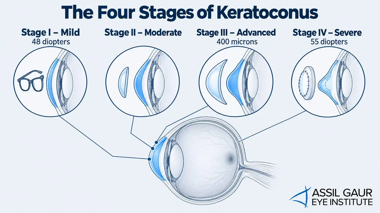
The Amsler-Krumeich staging system is the most widely used clinical framework for classifying keratoconus severity. It uses four stages based on corneal curvature (measured by keratometry), corneal thickness (measured by pachymetry), and corneal scarring. Understanding your stage is not just academic – it directly determines which treatment and vision correction pathways are available to you.
What are the stages of keratoconus?
The four Amsler-Krumeich stages classify keratoconus from mild (Stage I, curvature under 48 diopters, thickness above 500 microns) to severe (Stage IV, curvature above 55 diopters with scarring). Each stage reflects a different level of corneal structural compromise and directly determines which vision correction options remain viable. Patients in Stages I and II who achieve documented stability have the broadest range of surgical options, including ICL implantation.
Stage I (Mild): Corneal curvature below 48 diopters, no scarring, and central corneal thickness above 500 microns. Vision is often correctable to near-normal with glasses or soft contact lenses. This is the ideal stage for intervention – cross-linking here preserves the most options.
Stage II (Moderate): Curvature between 48 and 53 diopters, thickness between 400 and 500 microns. Glasses correction becomes less reliable; rigid gas-permeable or scleral contact lenses are typically needed. Surgical vision correction is still achievable after stabilization, but the corneal architecture is more compromised.
Stage III (Advanced): Curvature between 53 and 55 diopters, thickness below 400 microns. Scleral lenses remain the primary vision management tool. Implantable Contact Lenses (ICL) may still be an option depending on anterior chamber depth and residual corneal thickness, but the surgical planning becomes significantly more complex.
Stage IV (Severe): Curvature above 55 diopters, significant thinning, and often corneal scarring. At this stage, corneal transplantation may be the only path to functional vision. This is the outcome that early intervention is designed to prevent.
What Accelerates Keratoconus Progression (and the Habits That Make It Worse)
Eye rubbing is the single most modifiable risk factor for keratoconus progression. Repeated mechanical pressure on the cornea degrades collagen cross-links and accelerates thinning. This is not a minor habit – patients with atopy or allergic eye disease rub their eyes chronically, often without realizing it, and this population consistently shows faster progression rates. If you have seasonal allergies and keratoconus, managing the allergy is as important as managing the cornea.
UV-A light exposure is a secondary accelerant. The cornea’s collagen fibers are partially stabilized by natural UV exposure, which is why corneal cross-linking (CXL) uses UV-A light therapeutically – but chronic unprotected UV exposure in the context of already-compromised collagen architecture can worsen the structural imbalance. Wearing UV-blocking sunglasses is a practical, low-cost protective measure that every keratoconus patient should adopt immediately.
Genetic factors and a family history of keratoconus also predict faster progression. Research published in PubMed Central highlights that early and accurate detection of progression is particularly important for cost-effective use of corneal cross-linking – meaning the patients who benefit most from CXL are those identified before significant structural loss has occurred. Waiting for symptoms to worsen before seeking specialist evaluation is the most common and most costly mistake we see.
What causes keratoconus to get worse?
The primary drivers of accelerated keratoconus progression are chronic eye rubbing, atopic (allergic) eye disease, UV exposure without protective eyewear, and genetic predisposition. Younger patients progress faster because corneal collagen is more susceptible to structural change before natural age-related stiffening occurs. Patients who combine multiple risk factors – for example, a teenager with severe allergies who rubs their eyes daily – are at the highest risk of rapid progression and should be evaluated for cross-linking without delay.
How Specialists Monitor Progression: Corneal Tomography, Pachymetry, and What the Numbers Mean
Standard corneal topography maps the cornea’s front surface and is the most sensitive tool for detecting early keratoconus. But topography alone tells only part of the story. Corneal tomography, particularly Scheimpflug imaging (produced by systems like the Pentacam), creates a full three-dimensional map of both the anterior and posterior corneal surfaces, along with corneal thickness distribution across the entire cornea. This matters because posterior corneal changes often precede visible anterior changes – meaning tomography can detect progression that topography misses entirely.
Pachymetry measures corneal thickness at multiple points. A thinning cornea is the clearest structural sign of active progression. Serial pachymetry readings taken six to twelve months apart give clinicians a quantitative progression rate – not just a snapshot, but a trajectory. Keratometry readings (measuring corneal curvature in diopters) complete the picture, with steepening curvature confirming that the cone is advancing.

Corneal Cross-Linking: The Gold Standard for Halting Progression
Corneal cross-linking (CXL) is the only FDA-approved treatment proven to halt keratoconus progression. The procedure applies riboflavin (Vitamin B2) eye drops to the cornea, then activates them with UV-A light. The photochemical reaction creates new bonds between corneal collagen fibers, significantly increasing the cornea’s biomechanical stiffness and resistance to further deformation.
CXL does not restore vision already lost to keratoconus. It stops the clock. That distinction is critical: the earlier CXL is performed, the more corneal architecture is preserved, and the more vision correction options remain available afterward. A patient who undergoes CXL at Stage I with a corneal thickness of 520 microns has a fundamentally different surgical future than one who waits until Stage III with 390 microns remaining.
Clinical literature in CRST Europe notes that corneal topography remains the most sensitive method for detecting early keratoconus and following progression – reinforcing why specialist-level imaging, not a standard refraction, is the appropriate monitoring tool. At a general optometry practice, the imaging resolution and the clinical experience to interpret subtle progression signals may simply not be present.
Can keratoconus progression be stopped?
Yes – corneal cross-linking (CXL) is the established, FDA-approved method for halting keratoconus progression. By strengthening the bonds between corneal collagen fibers using riboflavin drops and UV-A light, CXL stabilizes the cornea and prevents further steepening and thinning. It does not reverse existing damage, which is why timing matters: CXL performed at Stage I or early Stage II preserves significantly more surgical options than CXL performed after advanced thinning.
Call now or book your appointment online.
What ‘Stable Keratoconus’ Means – and Why It’s the Gateway to Vision Correction Surgery
This is the concept most keratoconus content online fails to explain, and it matters most to patients who want freedom from glasses. “Stable keratoconus” means that serial measurements – topography, tomography, pachymetry, and keratometry – show no meaningful change over a defined monitoring period, typically one to two years. Stability is not the same as mild keratoconus. A patient can have moderately advanced keratoconus that has been stable for two years and still be a strong candidate for ICL implantation.
Stability is the clinical prerequisite for elective vision correction surgery in keratoconus patients. LASIK is contraindicated in keratoconus regardless of stability, because the laser ablation removes corneal tissue and further weakens an already compromised structure. But ICL – which adds a lens inside the eye without touching the cornea – is a fundamentally different procedure. For stable keratoconus patients with adequate anterior chamber depth, ICL can deliver dramatic vision improvement without any corneal risk.
Refractive Lens Exchange (RLE) is another pathway for appropriate candidates, particularly those in their forties or older whose natural lens is beginning to lose flexibility. The key point is that stability, achieved either through natural age-related stiffening or through CXL, reopens a door that many patients assume is permanently closed.
At what age does keratoconus stabilize?
Natural stabilization most commonly occurs between the mid-thirties and early forties, as the cornea’s collagen naturally stiffens with age. However, this timeline is not guaranteed – some patients continue to progress into their forties, particularly those with ongoing risk factors like eye rubbing or atopy. Patients who cannot wait for natural stabilization, or who are progressing rapidly in their twenties, should discuss CXL with a corneal specialist to accelerate the stabilization timeline and preserve their surgical options.
Can you have LASIK if you have keratoconus?
LASIK is contraindicated for patients with keratoconus, whether active or stable. The laser ablation required for LASIK removes corneal tissue, which further weakens a cornea that is already structurally compromised. Performing LASIK on a keratoconus cornea risks accelerating cone formation and causing post-LASIK ectasia, which can be severe and difficult to manage. ICL, RLE, and, in some cases, phakic IOL implantation are appropriate vision-correction alternatives for keratoconus patients who have achieved documented stability.
When LASIK Is Off the Table: How ICL and Other Premium Options Restore Vision for Keratoconus Patients
Being told you’re not a LASIK candidate is not the end of the vision correction conversation. It’s the beginning of a more specific one. At Assil Gaur Eye Institute, we regularly see patients who arrive having been told, “you can’t have LASIK,” with no further explanation of what they can have. That gap in guidance is unnecessary and, frankly, avoidable.
Implantable Contact Lenses (ICL) are the most common premium vision correction pathway for stable keratoconus patients. The ICL is placed between the iris and the natural lens, correcting refractive error without altering the cornea. For patients with high myopia and stable keratoconus, ICL can achieve results that rival LASIK outcomes in non-keratoconus patients. The procedure requires careful pre-operative measurement of anterior chamber depth and endothelial cell count, which is exactly the kind of precision evaluation our team performs as standard.
Consider the experience of a patient in their late twenties who came to us after being declined for LASIK at two other practices due to irregular topography. After a full tomographic workup confirmed stable Stage II keratoconus, and after a period of documented stability post-CXL, they were an excellent ICL candidate. The outcome: functional vision without glasses or contact lenses for the first time in their adult life. That kind of result requires both the diagnostic precision and surgical range a specialist practice provides.
For patients exploring premium intraocular lens options when they aren’t LASIK candidates, the range of available technologies has expanded significantly. The goal of freedom from glasses is achievable for many keratoconus patients – the path just requires more precise planning.
Dry eye management also plays a meaningful role in surgical candidacy and outcomes. Keratoconus patients who wear scleral contact lenses often develop secondary dry eye symptoms, and addressing this before surgery improves both the accuracy of pre-operative measurements and post-operative comfort. Understanding how nutritional factors influence corneal health and surgical outcomes is part of every pre-surgical conversation we have with patients.
Why Early Evaluation at a Specialist Practice like Assil Gaur Eye Institute Can Change Your Outcome
The difference between a general eye exam and a specialist corneal evaluation is not just the equipment – it’s the clinical pattern recognition that comes from seeing hundreds of keratoconus cases at every stage. At a busy general practice, subtle posterior corneal changes on a Scheimpflug scan may not trigger the same response they would at a practice where corneal conditions are a primary focus.
As noted in Modern OD, many patients are referred with moderate to advanced keratoconus that was missed or underappreciated at the primary care level – meaning the window for early CXL had already partially closed by the time they reached a specialist. Earlier referral, or proactive self-referral, changes that trajectory entirely.
If you’ve been told your prescription keeps changing, if you’ve been declined for LASIK, or if you have a family history of keratoconus, the most valuable thing you can do right now is get a specialist-level corneal evaluation.
At Assil Gaur Eye Institute, our team has performed over 100,000 vision correction surgeries and trained over 14,000 surgeons worldwide. We know what stable looks like, what progressive looks like, and exactly which pathway gives you the best chance at the vision you’re aiming for. Book your evaluation today and get a precise answer – not a general one.
Call now or book your appointment online.
Frequently Asked Questions
How fast does keratoconus progress?
Keratoconus progression varies significantly from patient to patient. During the teenage years and early twenties, the cornea can steepen by one to two diopters per year in rapidly progressing cases. In slower-progressing patients, changes may be barely measurable over several years. Age, genetic factors, eye-rubbing habits, and atopic (allergic) eye disease influence the rate. Serial corneal tomography measurements taken six to twelve months apart are the most reliable way to quantify an individual patient’s progression rate.
What happens if keratoconus is left untreated?
Without intervention, progressive keratoconus leads to increasing corneal steepening and thinning, worsening irregular astigmatism, and eventually vision that cannot be corrected with glasses. In advanced cases, corneal scarring develops, and the only remaining option may be a corneal transplant (penetrating keratoplasty or deep anterior lamellar keratoplasty). Untreated keratoconus also eliminates the possibility of elective vision correction procedures like ICL, which require a minimum level of corneal structural integrity. Early treatment with corneal cross-linking is specifically designed to prevent this cascade.

Written by The AGEI Educational Team

Medically Reviewed by Dr. Avneet Sodhi Gaur