Eye Strokes (Central Retinal Artery Occlusion)in Los Angeles
in Los Angeles
What is an eye stroke?
An eye stroke or anterior ischemic optic neuropathy is a dangerous and debilitating condition resulting from a lack of sufficient blood flow to the tissue in the front part of the eye’s optic nerve.
A stroke in the eye occurs when blood flow to the retina is interrupted due to a blockage. Several factors, including plaque or an infection in another part of the body, can cause the blockage. If the blood flow to the retina gets blocked, it robs the retinal cells of oxygen, and they begin to die off. This can result in blurred vision and permanent vision loss.
Immediate treatment is essential to prevent permanent vision damage and loss, as the retina can only survive for four to six hours.
Dr. P explains CRAO (also known as an eye stroke)
What are the symptoms of an eye stroke?
The hallmark symptom of central retinal artery occlusion (CRAO) is a sudden change in sight. It’s important to note that these symptoms typically occur suddenly, almost always in one eye, and without any accompanying pain.
Symptoms include:
- Flashes and floaters
- Distorted or blurry vision
- Blind or dark spots
- Vision changes that start slight but worsen over time
What causes eye strokes?
The blockage from an eye stroke occurs with plaque build-up or even an infection in the eye or another body part, like the heart or other arteries. This blockage is known as an embolism. According to the American Academy of Ophthalmology, men are at a higher risk of having an eye stroke than women.
Risk factors for eye stroke are similar to the risk factors for having a stroke in general, including:
- High blood pressure (hypertension)
- Diabetes
- Smoking
- Glaucoma
- High cholesterol (hyperlipidemia)
- Heart disease-plaque build-up in the blood vessels (Atherosclerosis)
- Individuals with a history of blood clots or migraines.
- Surviving a previous stroke
- Though uncommon, studies have shown that those with severe COVID can also develop eye strokes
Call now or book your appointment online.
How are eye strokes diagnosed?
Diagnosing an eye stroke typically involves a combination of the following:
Comprehensive eye exam
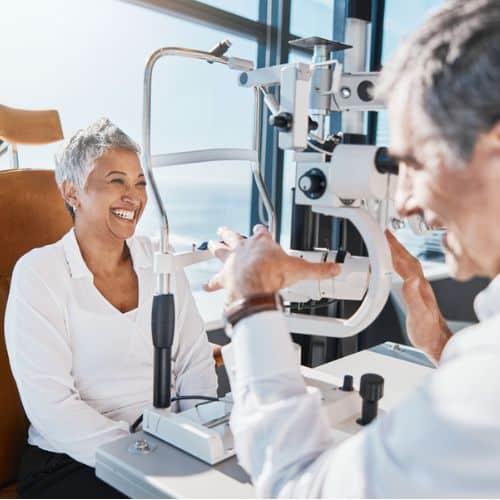
Your doctor will review your medical history and then begin an eye examination. An ophthalmologist will evaluate your visual acuity, examine the structures of the eye, and assess the blood flow to the retina. They may use instruments such as a slit lamp, fundoscopy (ophthalmoscopy), or a retinal camera to examine the back of the eye.
Imaging tests
Imaging tests are performed to confirm the diagnosis and evaluate the extent of the blockage. One common imaging technique is fluorescein angiography, in which a special dye is injected into a vein in the arm, and photographs are taken as the dye flows through the blood vessels in the retina. This test helps to identify areas of blockage or leakage.
Optical coherence tomography (OCT) is another imaging tool that uses light waves to create detailed cross-sectional images of the retina. It also provides information about the thickness of the retina and identifies any swelling or damage.
Blood work
In some cases, additional tests may be ordered, such as blood tests to assess for underlying conditions like diabetes or high cholesterol, or imaging of the carotid arteries to check for blockages that may have contributed to the eye stroke.
Is there treatment for eye strokes?
If you experience sudden vision loss or other symptoms of an eye stroke, it is crucial to seek immediate medical attention. Time is of the essence, and prompt evaluation is necessary to determine the best course of treatment.
The treatment for eye strokes, specifically retinal artery occlusion (CRAO), depends on the underlying cause and the timing of presentation. Some potential treatment options include:
Ocular massage
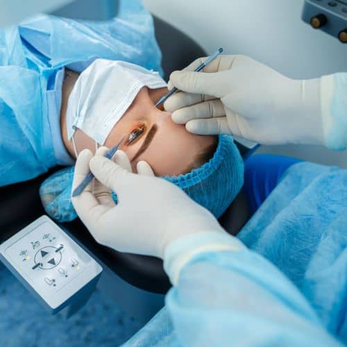
In some situations, a healthcare professional may perform a gentle massage of the affected eye to potentially dislodge an embolus and restore blood flow.
Hyperbaric Oxygen Therapy
This painless therapy involves breathing pure oxygen in a special chamber. The increased oxygen aids in preserving the energy supply of the retina
Medication
Medication may be used to dissolve the blood clot or reduce eye pressure.
The most important part of the treatment is an immediate stroke protocol workup, and patients are typically referred to the Emergency Room for a comprehensive evaluation of the heart, an ultrasound of the carotid arteries in the neck to identify possible emboli sources, and an urgent assessment of all stroke risk factors.
Eye strokes can increase your risk of a brain stroke. Patients will likely need long-term follow-up care to treat the problems (such as diabetes or cardiovascular disease) that may have contributed to the eye stroke.
Are there other types of eye strokes?
While central retinal artery occlusion (CRAO) is the most common type of eye stroke, there are others, including:
CRVO (Central Retinal Vein Occlusion)
CRVO, or Central Retinal Vein Occlusion, happens when the central vein of the retina becomes blocked or narrowed. This blockage prevents proper blood drainage from the retina, resulting in fluid accumulation and swelling.
The degree of loss of vision from CRVO depends on the extent of vein blockage and the size of the affected blood vessel. The most common risk factors for this condition are high blood pressure, diabetes, high cholesterol, and smoking.
BRVO (Branch Retinal Vein Occlusion)
Branch Retinal Vein Occlusion is similar to CRVO; it involves the blockage of one of the smaller veins (or branches) draining blood from the retina, not the central vein.
BRAO (Branch Retinal Artery Occlusion)
BRAO, like BRVO, affects the smaller branches, but BRAO affects the arteries that bring fresh blood to the retina. A blockage in one of these branches can cause a sudden loss of vision in a part of your visual field.
CRVO, CRAO, BRVO, and BRAO are all considered medical emergencies that require immediate medical attention.
Why choose Assil Gaur Eye Institute for eye stroke treatment
The AGEI staff includes a highly skilled retina specialist, Dr. Svetlana Pilyugina, or “Dr. P,” as she is known to her patients. Dr. Pilyugina is a Stanford-trained ophthalmologist with fellowship training and board certification in diseases and surgery of the vitreous and retina.
Dr. P has extensive experience treating macular degeneration, diabetic eye disease, retinal tears and detachment, flashes, floaters, and complex retinal conditions.
Apart from her eye care experience, Dr. P is recognized for her dedication to her patients and her focus on personalized service. That is why she limits the number of patients she sees daily in the clinic, allowing her to dedicate sufficient time for evaluation and consultation with each patient to meet her high standard of care.
The Assil Gaur Eye Institute team of ophthalmologists and optometrists are conveniently located for patients throughout Southern California and the Los Angeles area in or near Beverly Hills, Santa Monica, West Los Angeles, West Hollywood, Culver City, Hollywood, Venice, Marina del Rey, Malibu, Manhattan Beach, and Downtown Los Angeles.
Read real patient stories about retinal conditions.
-
Central retinal vein occlusion
https://www.nei.nih.gov/learn-about-eye-health/eye-conditions-and-diseases/central-retinal-vein-occlusion-crvo -
What Is Central Retinal Vein Occlusion (CRVO)?
https://www.aao.org/eye-health/diseases/what-is-central-retinal-vein-occlusion -
aRetinal Artery and Vein Occlusions Successfully Treated with Hyperbaric Oxygen
https://www.ncbi.nlm.nih.gov/pmc/articles/PMC6861035/ -
The Incidence of Central Retinal Artery Occlusion in Olmsted County, Minnesota
https://www.ncbi.nlm.nih.gov/pmc/articles/PMC3326414/ -
Retinal manifestations in patients with SARS-CoV-2 infection and pathogenetic implications: a systematic review
https://www.ncbi.nlm.nih.gov/pmc/articles/PMC8356207 -
4 Ways COVID Leaves Its Mark on the Eye
https://www.aao.org/eye-health/tips-prevention/covid-damages-retina-eye-strokepinkeye


16,884 TOTAL 1ST PARTY REVIEWS / 4.9 OUT OF 5 STARS





social media for more