Nevus (Eye Freckle) Removal
Expert Conjunctival Nevus (Eye Freckle) Removal in Los Angeles
What is a conjunctival nevus?
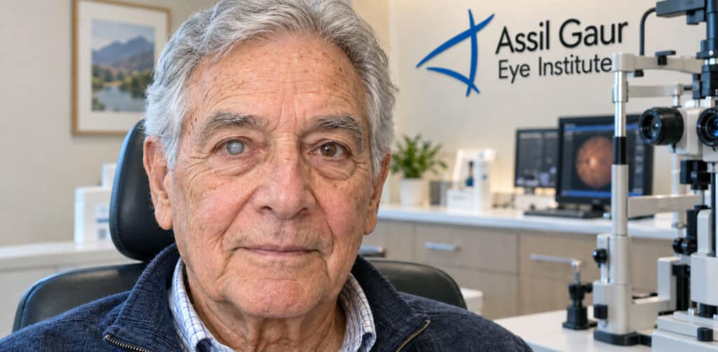
A conjunctival nevus is a spot that looks like a freckle or mole and is usually found on the transparent tissue covering the eye. Nevi (the plural of nevus) can vary from dark brown to yellow, and over time, these spots might become darker or lighter. They are typically located near the iris, which is the colored part of the eye.
Nevi are composed of pigment cells called melanocytes that give color to your skin, hair, and eyes. When multiple melanocytes cluster together, they form a nevus.
They are commonly found in Caucasians because light eyes, fair skin, and people who burn easily when exposed to the sun are also risk factors.

A conjunctival nevus is a benign melanocytic lesion that forms on the ocular surface, the thin, transparent tissue covering the white of the eye. Most are congenital and present at birth, though acquired nevi can appear later in life. For many patients, the first question is not whether to monitor it, but whether to remove it. At Assil Gaur Eye Institute (AGEI), the answer is yes, through a minimally invasive technique pioneered by Dr. Kerry Assil that leaves no visible trace.
What causes eye freckles?
Conjunctival nevi arise from melanocytes, the pigment-producing cells embedded in the conjunctival tissue that lines the white of your eye and the inner surface of your eyelids. These cells produce melanin, the same pigment responsible for skin and hair color, and when they cluster together abnormally, a nevus forms. Think of it as a freckle on the skin, but located on the surface of the eye instead.
Nevi fall into two broad categories. Congenital nevi are present at birth and result from melanocyte clustering during fetal development. Acquired nevi, which are far more common, typically appear during childhood or adolescence and may gradually darken or enlarge during hormonal shifts such as puberty or pregnancy. This is completely normal behavior for a benign nevus, though any sudden change in size, color, or shape always warrants a prompt evaluation.
Large-scale studies have linked prolonged UV exposure to nevus development, and the American Academy of Ophthalmology (AAO) confirms that ultraviolet light may play a meaningful role in their formation. People with lighter skin tones and lighter eye pigmentation carry a somewhat higher risk, likely because lower baseline melanin levels leave the conjunctival tissue more vulnerable to UV-related cellular changes. The AAO also clarifies that a family history of nevi does not meaningfully increase your personal risk, which surprises many patients who assume the condition runs in families the way other eye conditions do.
Environmental habits matter too. Extended time outdoors without UV-protective sunglasses, particularly in high-altitude or high-reflection environments like snow or open water, increases cumulative UV exposure to the ocular surface. Wearing quality wraparound sunglasses is one of the simplest steps you can take to protect your conjunctival tissue over a lifetime.
How worried should I be about an eye freckle?
Most conjunctival nevi are benign and remain stable throughout a person’s lifetime. That said, understanding the difference between a harmless nevus and something more serious is exactly why periodic monitoring matters.
Conjunctival melanoma is a malignancy that arises from the ocular surface, specifically the conjunctiva, the thin transparent tissue covering the white of the eye. It is entirely separate from uveal melanoma or choroidal melanoma, which originate deeper in the eye from the pigmented layer beneath the retina. The two have different risk profiles and treatment pathways, and should not be confused. Conjunctival melanoma is rare: published clinical studies estimate that malignant transformation occurs in fewer than 1 in 200 conjunctival nevi, a rate of roughly 0.5 percent over a lifetime of observation.
A nevus present since childhood is almost always benign. One that appears for the first time in adulthood, or that changes in size, color, or vascularity, warrants closer attention, much like a new or changing skin mole would prompt a visit to a dermatologist. Even then, the overwhelming majority of adult-onset conjunctival nevi are still harmless. The key is not to panic, but to monitor consistently. Most ophthalmologists recommend a baseline photograph at diagnosis, a follow-up exam at six months, and then annual or biennial reviews depending on the nevus characteristics. Any lesion that grows, darkens, develops new blood vessels, or loses its cystic appearance should be evaluated promptly rather than waiting for the next scheduled visit.
Periodic eye exams with a specialist who can document and compare changes over time remain the most reliable safeguard against missing a meaningful shift in a conjunctival nevus.
When should you see your eye doctor about an eye freckle?
An ophthalmologist should evaluate any new pigmented lesion on the eye surface. Seek prompt care if you notice:
Blurry vision or other vision changes
Eye floaters or flashing lights
Eye pain or discomfort
Changes in the size or color of the freckle
These symptoms can signal a serious underlying problem. Choroidal nevi can occasionally leak fluid or trigger abnormal blood vessel growth, and if left unaddressed, either complication can progress to retinal detachment and permanent vision loss.
What are the treatment options for conjunctival nevus?
Treatment exists on a spectrum, and the right path depends on the nevus itself – its size, depth, location, and whether it shows any signs of change. Our physicians examine the front and interior structures of the eye, including the retina, macula, and optic nerve, and use photodocumentation to track any changes over time. Using the ABCDE framework, your doctor monitors for:
Asymmetrical shape
Irregular borders
Color changes or multiple colors
Diameter that has increased
Evolution of the nevus appearance over time
A baseline visit is typically followed by a six-month recheck. If the nevus is stable over one to two years, the risk of malignant transformation is very low, though annual monitoring remains advisable.
When it comes to actual treatment, three distinct clinical pathways exist, and understanding the difference matters.
Observation with photodocumentation is the first-line approach for stable, small nevi that show no concerning features. Serial photographs allow your physician to detect even subtle changes between visits without any intervention.
Argon laser ablation is an option for superficial, pigmented lesions that sit close to the surface of the conjunctiva. The laser targets pigmented cells precisely, reducing the nevus without an incision. It works best on flat, well-defined nevi and is generally not appropriate for deeper or larger growths.
Surgical excision with pathology review is the gold standard when a nevus shows growth, color change, or other warning signs, or when a patient elects removal for cosmetic reasons. After removal, the tissue is sent to a pathologist for optin a definitive answer on whether any cells were atypical. At Assil Gaur Eye Institute, Dr. Assil has pioneered a minimally invasive thermal brushing technique that removes the nevus with exceptional precision while minimizing disruption to the surrounding conjunctival tissue, supporting a smoother recovery and enviable outcomes.
Choosing between these options is not a one-size-fits-all decision. Your AGEI physician will walk you through the clinical picture specific to your eye and help you understand which approach aligns with both your health needs and your goals.
SEE LIFE THROUGH OUR PATIENT'S EYES
REAL NEVUS REMOVAL PATIENT STORIES



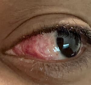

Conjunctival nevus removal: the procedure

AGEI removes conjunctival nevi for two reasons: suspected malignancy and cosmetic concerns.
Medically indicated excisional biopsy: When cancer is suspected, the pigmented lesion is removed surgically with clean surgical margins and sent to a pathology lab to assess for malignant cells. This excisional biopsy is the clinical standard and may be covered by insurance when medically necessary.
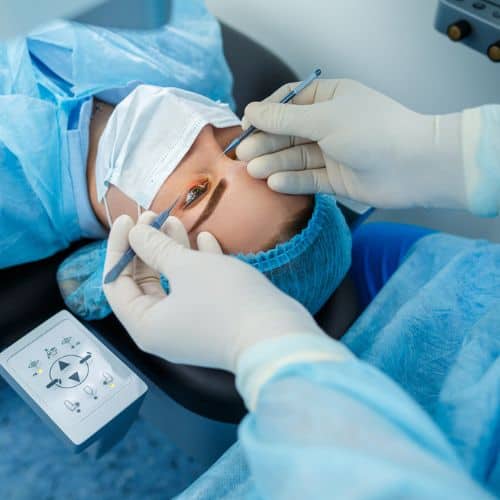
Cosmetic removal using thermal brushing: Dr. Kerry Assil pioneered a proprietary thermal cautery technique that gently extracts pigment from the conjunctival surface without the tissue disruption of traditional surgical excision. Unlike argon laser ablation, which targets pigment with focused light energy, thermal brushing allows precise, layer-by-layer removal with minimal involvement of surrounding tissue. The procedure is performed in-office, typically completed in under 30 minutes from consultation to completion, and leaves no visible trace of the prior nevus. Recovery is quick and largely painless.
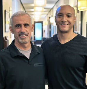
NBA Hall of Famer Dwyane Wade chose AGEI and Dr. Assil for his conjunctival nevus removal, reflecting the trust world-class athletes place in this technique.
Request Your Consultation
Call now or book your appointment online.
Am I a good candidate for conjunctival nevus removal?
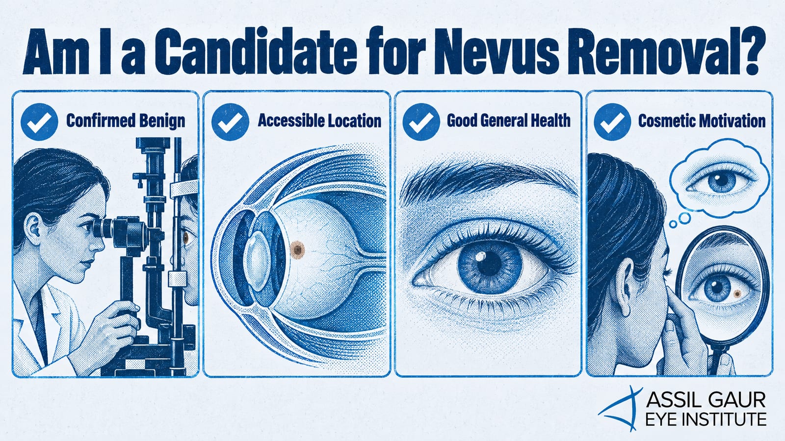
Most patients with a stable, benign conjunctival nevus are candidates for cosmetic removal. You are likely a strong candidate if:
Your nevus has been evaluated and confirmed benign by an ophthalmologist
The lesion is located on the accessible conjunctival or limbal surface
You are in good general health with no active ocular infection
Your primary motivation is cosmetic, and you understand the procedure is elective
Patients with suspicious features, rapid growth, or irregular borders may be directed toward excisional biopsy first. A thorough consultation at AGEI determines the right approach for your specific lesion.
Recovery and aftercare
After cosmetic removal using Dr. Assil's thermal brushing technique, most patients experience mild redness or irritation for a few days. Prescription eye drops manage inflammation and reduce infection risk. Patients typically return to normal activities within a week, with full ocular surface healing complete within two to four weeks. Follow-up visits confirm clean healing and allow your physician to document the treated site.
Cost and insurance coverage
Cosmetic conjunctival nevus removal is considered an elective procedure and is not covered by insurance. Patients should expect to pay out of pocket, and AGEI's team can provide a clear cost estimate at your consultation. When removal is medically indicated, such as an excisional biopsy for a suspicious lesion, insurance may cover the procedure. Our staff will verify your benefits and walk you through your options before any treatment begins.
Why choose AGEI for your eye freckle removal in Los Angeles
When it comes to conjunctival nevus removal, experience and technique matter enormously. AGEI's physicians have performed thousands of conjunctival nevus removals, and the institute is nationally recognized for Dr. Assil's proprietary thermal brushing technique, a minimally invasive approach that gently removes the nevus tissue with precision while preserving the surrounding conjunctiva. This exclusive method offers a meaningful advantage over traditional surgical excision, which carries a higher risk of scarring, and argon laser ablation, which can leave residual pigmentation. The thermal brushing technique delivers clean, cosmetically superior results with a faster, more comfortable recovery.
AGEI's reputation for conjunctival nevus removal extends well beyond Los Angeles. Patients travel from across the country and around the world to have Dr. Assil perform this procedure, including high-profile cases that have brought national attention to the institute's world-class outcomes. Los Angeles Magazine has named AGEI among the region's top ophthalmology practices year after year, and the institute's surgeons have earned the trust of elite athletes and celebrities seeking the very best in precision eye care.
Patients interested in related ocular surface concerns can also explore AGEI's specialized care for Pterygium (Surfer's Eye), a condition that shares overlapping UV exposure risk factors with conjunctival nevi, or browse the full range of Oculoplastics and Aesthetics procedures available at the institute.
Read real patient stories about nevus removal surgery.
If you have noticed a spot on the white of your eye and want a definitive answer on whether it should be monitored or removed, AGEI is the place to start. Schedule your complimentary evaluation by calling 866-945-2745 or requesting an appointment online. Our world-class team, led by the surgeon who pioneered the thermal brushing technique, is ready to deliver the nationally recognized, enviable outcomes you deserve.
We serve patients throughout Southern California from locations convenient to Beverly Hills, Santa Monica, West Los Angeles, West Hollywood, Culver City, Hollywood, Venice, Marina del Rey, Malibu, Manhattan Beach, and Downtown Los Angeles.
Request Your Consultation
Call now or book your appointment online.
Eye Freckle FAQs
Are there different types of nevi?
There are different types of nevi based on where the nevus is located.
- Conjunctival nevus
This type of eye freckle is the conjunctiva, the transparent film covering the front of your eyes. It usually shows up because it appears on the sclera or the white part of your eye. Conjunctival nevi are the common type of nevi. -
Iris nevus and iris freckle
An iris nevus and iris freckles appear in the iris. An iris freckle is smaller than an iris nevus. However, an iris nevus goes deeper into the iris and sometimes can pull the pupil to the side. -
Choroidal nevus
A choroidal nevus occurs in the choroid, the layer of tissue beneath the retina in the back of the eye. The choroid is part of the uvea, the pigmented part of your eye, and includes the iris. Both choroidal nevi and iris nevi are forms of uveal nevi.
What is recovery like after eye freckle removal, and how soon can I return to normal activities?
Recovery is typically quick and comfortable, with no stitches and most patients back to their normal routine within a few days. Mild redness, a gritty feeling, or light watering in the first several days is normal, and your surgeon will give simple aftercare instructions like avoiding eye rubbing and swimming for a short period
Sources
-
Conjunctival Nevi: Clinical Features and Natural Course in 410 Consecutive Patients
https://doi.org/10.1001/ARCHOPHT.122.2.167 -
Conjunctival naevi in Denmark 1960-1980. A 21-year follow-up study
https://www.ncbi.nlm.nih.gov/pubmed/8883545 -
Conjunctival lesions in adults. A clinical and histopathologic review
https://www.ncbi.nlm.nih.gov/pubmed/3301209 -
Subepithelial conjunctival nevus with atypia: Expanding our understanding of a challenging diagnosis
https://www.sciencedirect.com/science/article/pii/S2214330018300300

Written by The AGEI Educational Team

Medically Reviewed by Dr. Kerry Assil
No form shortcode found.