3mm Pupils: Normal Range, Causes & When to Worry
Understanding constricted pupils (pinpoint pupils)
Miosis refers to abnormally small, constricted pupils. Your pupils – the black openings at the center of each iris – naturally constrict and expand in response to light and focal distance. In bright light, a normal pupil typically measures between 2mm and 4mm, so 3mm pupils generally fall within the expected range under well-lit conditions. A 3mm measurement sits at the smaller end of the normal range in bright conditions, but the same measurement in a dim room, or paired with other symptoms, can signal a neurological or toxic emergency. If you’re concerned about your pupil size, the ophthalmologists at Assil Gaur Eye Institute (AGEI) can provide a thorough evaluation.
What are normal pupil size changes?
On average, pupils measure 2-4 millimeters in bright light and 4-8 millimeters in dim light. A measurement of 3mm pupils in a well-lit environment sits at the smaller end of that normal range – perfectly acceptable in bright conditions, and not a cause for concern on its own. A 3mm pupil in a well-lit room is therefore within the normal range. That same 3mm reading in darkness, however, suggests the pupil is failing to dilate as expected – a finding that warrants clinical attention.
The pupillary light reflex governs this response: the parasympathetic nervous system drives the iris sphincter muscle to constrict the pupil in bright light, while the sympathetic nervous system relaxes it to allow dilation in the dark. Focusing on a near object also triggers constriction; shifting gaze to a distant target allows the pupil to widen. Context matters enormously here. A 3mm reading that stays fixed regardless of lighting conditions, or that differs noticeably between your two eyes, tells a very different clinical story than one that simply reflects a well-lit room. Size alone is only part of the picture – reactivity and symmetry are equally important signals that a world-class eye care specialist will evaluate alongside the raw millimeter measurement. Disruption anywhere along these pathways – from the brainstem to the iris itself – can produce abnormal pupil size or reactivity.
When are 3mm pupils a concern?
Now that you understand how pupils naturally shift in size throughout the day, you might wonder where 3mm falls on the spectrum. The short answer is that 3mm pupils are generally considered normal, sitting comfortably within the typical bright-light range of 2 to 4 millimeters. In most cases, if your pupils measure around 3mm in a well-lit room, nothing clinically alarming is going on.
That said, context matters enormously. A 3mm pupil becomes a concern when the lighting conditions don’t match what you would expect. For example, if your pupils remain fixed at 3mm in a dim environment where they should naturally dilate to 4- 8 mm, that lack of response is worth paying attention to. Pupils that fail to react to changing light levels may signal an underlying issue with the nervous system or the eye itself.
Another red flag is asymmetry. If one pupil measures 3mm while the other is noticeably larger or smaller, that difference, known as anisocoria, can point to conditions ranging from Horner’s syndrome to more serious neurological events. A size difference of more than 1mm between pupils warrants a prompt evaluation.
Certain medications, including pilocarpine eye drops used to treat glaucoma, can also keep pupils constricted regardless of light. If you take any eye or systemic medications and notice your pupils seem unusually small or unresponsive, tell your eye care provider.
Call now or book your appointment online.
Potential causes for pupil constriction
Beyond normal physiologic responses, several medical conditions, medications, and toxic exposures can make pupils abnormally small or fixed. Each cause below is anchored to its clinical context, including the millimeter thresholds that separate a routine finding from a red flag worth investigating.
Horner’s syndrome
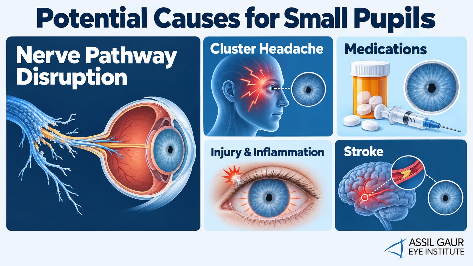
Also known as oculosympathetic palsy, Horner’s syndrome disrupts the sympathetic pathway from the brainstem to the eye. The affected pupil typically measures 1- 2 mm smaller than its partner, producing anisocoria that is most obvious in dim light. Associated findings include a droopy upper eyelid and lighter iris color on the affected side. Because Horner’s syndrome can be caused by a lung tumor, carotid artery dissection, or other serious pathology, new-onset cases require urgent imaging.
Cluster headache with small pupils
Cluster headaches can produce a transient Horner’s-like picture – unilateral miosis and ptosis – during an attack. Each episode typically lasts 15-90 minutes and can recur up to eight times in 24 hours. Because the presentation mimics true Horner’s syndrome, an ophthalmologist evaluation is important to rule out a structural cause before attributing small pupils to headache alone.
What drugs make your pupils small?
Several drug classes cause miosis by stimulating the parasympathetic system or suppressing sympathetic tone:
Opioids – Codeine, oxycodone, and fentanyl produce bilateral pinpoint pupils; in overdose, pupils may fix at 1- 2 mm and become non-reactive to light, a toxicology emergency.
Cholinergic agents – Organophosphate pesticides and nerve agents flood the parasympathetic system, causing profound miosis alongside excessive secretions and muscle weakness. Pupils can drop well below 3mm and become nearly fixed, which is a life-threatening emergency requiring immediate medical intervention.
Cholinesterase inhibitors – Medications used for Alzheimer’s disease (e.g., donepezil) can produce mild miosis through the same mechanism. Unlike organophosphate poisoning, miosis from therapeutic doses is typically subtle, often keeping pupils in the 2-3 mm range without other alarming signs.
Diazepam – This anti-anxiety sedative can constrict pupils as part of its CNS depressant effect.
Clonidine – This antihypertensive reduces sympathetic outflow, allowing parasympathetic tone to dominate.
Antihistamines – Over-the-counter options like Benadryl occasionally cause miosis, particularly in higher doses.
Pilocarpine eye drops – Used to treat glaucoma, pilocarpine directly stimulates the iris sphincter muscle and reliably produces miosis.
Clinically, the key distinction is whether miosis is bilateral and fixed or simply mild, reactive constriction. Bilateral pinpoint pupils that do not respond to light, especially when paired with altered consciousness, slowed breathing, or excessive secretions, point toward opioid overdose or cholinergic toxicity and demand emergency care. If you notice persistent or unexplained changes in your pupil size, a world-class evaluation at AGEI can help rule out underlying conditions and guide next steps.
Injury
Traumatic head injury can cause pupils to constrict through nerve damage or internal hemorrhage. A fixed, non-reactive pupil after head trauma is a neurological red flag requiring immediate emergency evaluation.
Eye inflammation
Conditions like iritis or uveitis involve inflammation inside the eye that can trigger reflex miosis in response to pain. Adie’s pupil, a benign condition caused by damage to the ciliary ganglion, produces a pupil that is poorly reactive to light and may appear asymmetric.
Stroke
A brainstem stroke can cause bilaterally pinpoint, non-reactive pupils – one of the most ominous signs in clinical medicine. Additional symptoms include double vision, dizziness, slurred speech, and severe balance problems. When pupils drop well below the normal 2- 4 mm range and appear fixed and non-reactive alongside any of these neurological warning signs, this combination points to a life-threatening emergency that demands an immediate 911 call rather than a wait-and-see approach. If a stroke is suspected, call 911 immediately; every minute of delay increases permanent neurological damage.
When to seek emergency care
Seek emergency care immediately if small or unequal pupils appear alongside any of the following:
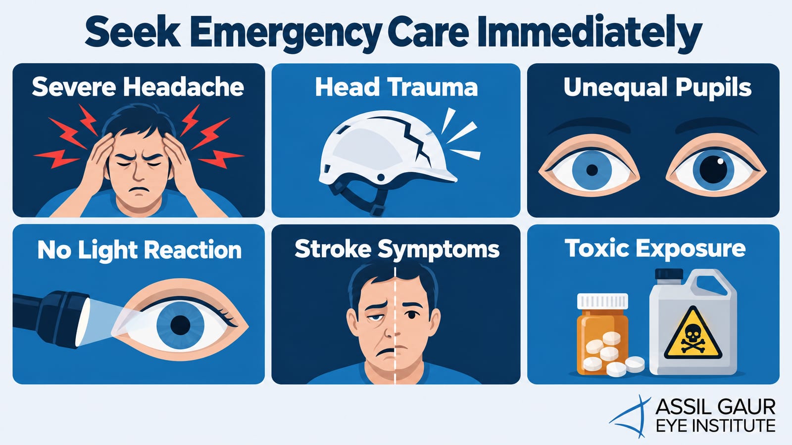
Sudden severe headache, especially described as “the worst of my life”
Head trauma or loss of consciousness
One pupil significantly larger or smaller than the other (anisocoria of 1mm or more in normal light)
Pupils that do not react to a bright light source
Slurred speech, facial drooping, arm weakness, or sudden vision loss
Known or suspected opioid or organophosphate exposure
These combinations can indicate stroke, herniation, opioid overdose, or cholinergic toxidrome – all of which are time-sensitive emergencies where rapid treatment is the difference between full recovery and permanent harm.
What a doctor will check
When you present with abnormal pupil size, an ophthalmologist or emergency physician will assess pupil diameter in both bright and dim light, test the pupillary light reflex in each eye, and look for anisocoria. They will review your medications and any toxic exposures, perform a neurological screen, and order imaging – typically a CT or MRI of the brain and brainstem – if a structural cause is suspected. Pharmacologic testing with dilute pilocarpine or apraclonidine drops can help confirm Horner’s syndrome or Adie’s pupil when the clinical picture is ambiguous.
Why trust Assil Gaur Eye Institute?
AGEI has assembled a team of nationally recognized ophthalmologists who deliver world-class care for the full spectrum of eye conditions, including pupil abnormalities that may signal serious neurological or ocular disease. Patients consistently say, “I knew I was going to the very best.” Whether your pupils measure 3mm, are smaller, or are unequal, our specialists have the advanced diagnostic tools and clinical expertise to identify the cause and guide your next steps with precision. Call (866) 945-2745 or schedule an online consultation.
Call now or book your appointment online.

Written by The AGEI Educational Team

Medically Reviewed by Dr. John May