LPI for Narrow-Angle Glaucoma
Your eye pressure is elevated, your angles are narrow, and your ophthalmologist just told you that you need a laser procedure – before you’ve experienced a single symptom of vision loss. That’s a disorienting place to be. You feel fine. Your vision seems normal. And yet the recommendation is urgent.
We hear this from patients at Assil Gaur Eye Institute all the time. The instinct is to wait, to get a second opinion, or to wonder whether the procedure will sideline you from work, training, or performance. Those are exactly the right questions to ask – and the ones most generic glaucoma resources never answer.
This guide explains what laser peripheral iridotomy (LPI) is, why acting before symptoms appear is the single most important decision you can make for your long-term vision, and what the procedure actually means for your daily life. You’ll learn how the procedure works mechanically, who the right candidates are, and what recovery looks like for patients who cannot afford visual downtime. If you’re already exploring your options, our world-class glaucoma care at Assil Gaur Eye Institute starts with a thorough evaluation before you decide on treatment.
What Is Laser Peripheral Iridotomy – and Why Would You Need It Before Noticing Any Symptoms?
Laser peripheral iridotomy is a brief, in-office laser procedure that creates a small opening in the peripheral iris – the colored part of your eye – to improve the flow of aqueous humor and reduce the risk of a dangerous spike in intraocular pressure (IOP). According to the Glaucoma Research Foundation, LPI has been used since 1984 to treat and prevent angle-closure glaucoma.
You can need this procedure without feeling anything because of your eye’s structure. Some eyes are simply built with a narrower anterior chamber angle – the space where the peripheral cornea and iris meet. That anatomy alone doesn’t cause symptoms. But it creates a condition where the iris can shift forward and block the trabecular meshwork, the drainage structure that regulates fluid outflow. When that blockage occurs suddenly, eye pressure can spike to dangerous levels within hours, causing permanent optic nerve damage before you’ve had time to react.
LPI eliminates that risk by creating an alternative pathway for aqueous humor to flow. The small hole in the iris bypasses the pupillary block mechanism entirely, keeping the drainage angle open even if the iris shifts. For patients with narrow angles who are otherwise asymptomatic, this is a preventive intervention – not a response to damage that has already occurred.

How Narrow Angles and Angle-Closure Glaucoma Put Your Optic Nerve at Risk
Think of the eye’s drainage system the way the Glaucoma Research Foundation describes it: like a sink. Aqueous humor flows in from the faucet and exits through the drain. In a healthy eye, that balance keeps IOP stable. In a narrow-angle eye, the iris sits too close to the trabecular meshwork and partially obstructs the drain. If the iris moves forward enough to seal the angle completely, the drain closes – and pressure builds rapidly.
The optic nerve is the structure most vulnerable to that pressure. It transmits visual information from the retina to the brain, and elevated IOP compresses the nerve fibers at the back of the eye. Unlike most tissues in the body, optic nerve fibers do not regenerate. Damage is permanent. This is why glaucoma is the leading cause of irreversible blindness worldwide, and why the standard of care for narrow-angle patients is to intervene before an acute attack occurs – not after.
Open-angle glaucoma, by contrast, involves a different mechanism: the trabecular meshwork becomes less efficient over time, causing a gradual, painless rise in pressure. LPI is not the treatment for open-angle glaucoma. It is specifically indicated for angle-closure mechanisms, where the iris itself is the obstruction.
How the LPI Procedure Works: A Step-by-Step Look at What Happens in the Laser Suite
The procedure is performed in the office, typically takes 5 to 10 minutes per eye, and requires no incisions, injections, or general anesthesia. Here is exactly what happens.
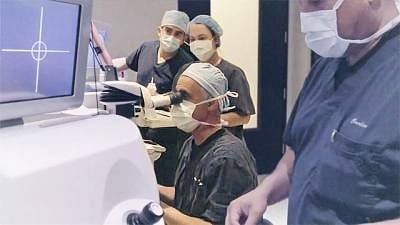
First, your surgeon applies a topical anesthetic drop to numb the eye surface. A special contact lens is placed on the eye to stabilize it and focus the laser precisely on the peripheral iris – the area closest to the edge, where the hole will be least visible and least likely to affect your vision. The YAG laser then delivers a series of precisely calibrated pulses to create a small, full-thickness opening through the iris tissue.
The entire energy delivery takes seconds. You may see a brief flash of light and feel a mild pressure sensation with each pulse, but the procedure is not painful. Once the opening is confirmed, the lens is removed, a pressure-lowering drop is applied, and you are monitored briefly before going home. The American Academy of Ophthalmology notes that most patients can resume normal activities the same day, with some light sensitivity and mild blurring that typically resolves within 24 hours.
Who Is a Candidate for Laser Peripheral Iridotomy? Understanding Patient Selection
Not every patient with elevated eye pressure needs LPI. The procedure is specifically indicated when the mechanism of pressure elevation – or the risk of it – involves the anterior chamber angle. Your surgeon will distinguish between narrow-angle risk and open-angle glaucoma before recommending LPI, because the two conditions require different interventions.
Consider the case of Dr. Klaus Ito, an optometrist who came to AGEI wearing hard contact lenses and experiencing chronic dry eyes during patient examinations. He wanted vision correction, but during his consultation with Dr. Avneet Gaur, they discovered and repaired a previously undetected retinal tear before any laser procedure. He then underwent Eagle Vision LASIK and achieved 20/10 vision in each eye. His case illustrates a principle we apply to every patient: thorough evaluation before treatment, not treatment before evaluation.
For LPI specifically, the evaluation includes gonioscopy to directly visualize the anterior chamber angle, IOP measurement, optic nerve assessment, and often optical coherence tomography (OCT) imaging to establish a baseline. As patient Julie H. noted after her experience at AGEI: “Dr. May had the technician take high-tech scans of my eyes, and six months later compared new scans with the originals. With such careful procedures, he was able to determine my exact needs.” That same precision governs every glaucoma evaluation we perform.
What to Expect Before, During, and After Your LPI Procedure
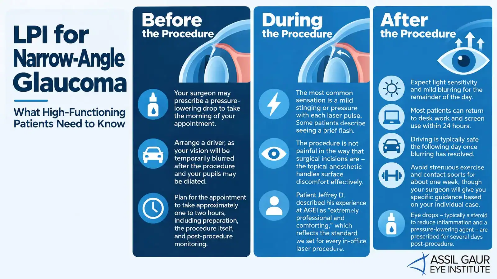
Before the procedure, your surgeon may prescribe a pressure-lowering drop to take the morning of your appointment. Arrange a driver, as your vision will be temporarily blurred after the procedure and your pupils may be dilated. Plan for the appointment to take approximately one to two hours, including preparation, the procedure itself, and post-procedure monitoring.
During the procedure, the most common sensation is a mild stinging or pressure with each laser pulse. Some patients describe seeing a brief flash. The procedure is not painful in the way that surgical incisions are – the topical anesthetic handles surface discomfort effectively. Patient Jeffrey D. described his experience at AGEI as “extremely professional and comforting,” which reflects the standard we set for every in-office laser procedure.
After LPI, expect light sensitivity and mild blurring for the remainder of the day. Most patients can return to desk work and screen use within 24 hours. Driving is typically safe the following day once blurring has resolved. Avoid strenuous exercise and contact sports for about one week, though your surgeon will give you specific guidance based on your individual case. Eye drops – typically a steroid to reduce inflammation and a pressure-lowering agent – are prescribed for several days post-procedure.
Is laser peripheral iridotomy painful?
LPI is not a painful procedure for the vast majority of patients. Topical anesthetic drops numb the eye surface before the laser contact lens is placed, and the laser pulses themselves last only fractions of a second. Most patients describe mild pressure or a brief stinging sensation with each pulse. Patients with darker, denser iris pigmentation may require more laser energy to create the opening, which can slightly increase the sensation, but this remains well within the range of tolerable discomfort for nearly all patients.
What is the recovery time after laser peripheral iridotomy?
Functional recovery – meaning the ability to return to screen work, reading, and light daily activity – typically occurs within 24 to 48 hours. Light sensitivity and mild blurring on the day of the procedure are normal. For patients with physically demanding schedules, including athletes and performers, we recommend avoiding high-intensity exercise, contact sports, and environments with significant dust or debris for approximately one week. A follow-up appointment is scheduled within two to four weeks to confirm that IOP has responded appropriately and that the iridotomy remains patent (open).
Laser Peripheral Iridotomy Cost, Insurance, and What ‘World-Class’ Care Actually Includes
LPI is a medically indicated procedure for patients with narrow-angle glaucoma or angle-closure risk, which means it is typically covered by medical insurance – including Medicare – when properly documented. This distinguishes it from elective refractive procedures. Your practice’s billing team should verify your specific coverage before the procedure and handle any prior authorization requirements.
Call now or book your appointment online.
LPI vs. Other Glaucoma Treatments: Where Laser Peripheral Iridotomy Fits in Your Care Plan
LPI is the first-line treatment for angle-closure glaucoma and narrow-angle risk, but it is not the only tool in a glaucoma specialist’s arsenal – and it does not work in isolation for every patient. Understanding where it fits in the broader treatment continuum matters, especially if your pressure remains elevated after the procedure.
Laser trabeculoplasty (LTP) is a different laser procedure used primarily for open-angle glaucoma. While LPI creates a hole in the iris to bypass a pupillary block, LTP applies laser energy to the trabecular meshwork to improve drainage efficiency. The two procedures address different mechanisms and are not interchangeable. As the BrightFocus Foundation notes, laser trabeculoplasty is often used when medications are unlikely to work or produce intolerable side effects in open-angle patients, while LPI is the preferred first intervention in angle-closure cases.
If LPI successfully opens the angle but IOP remains elevated, the next step is typically topical glaucoma medications – prostaglandin analogs, beta-blockers, or carbonic anhydrase inhibitors – to reduce aqueous humor production or increase outflow. If medications are insufficient, laser trabeculoplasty or surgical options, including trabeculectomy (a filtration surgery that creates a new drainage channel), may be considered. The key point is that LPI is a decision point, not a destination. A glaucoma specialist will monitor your IOP and optic nerve at regular intervals after LPI to determine whether you need additional intervention.
What happens if laser peripheral iridotomy doesn’t work?
If LPI successfully opens the anterior chamber angle but IOP remains elevated, it indicates that the trabecular meshwork itself has sustained damage from chronic angle narrowing – meaning the drainage system needs additional support beyond the angle being opened. In this scenario, your glaucoma specialist will typically add topical IOP-lowering medications as the next step. If pressure remains uncontrolled despite drops, laser trabeculoplasty or surgical intervention such as trabeculectomy or a minimally invasive glaucoma surgery (MIGS) procedure may be recommended. In rare cases, the iridotomy opening may close over time and require retreatment. This is why post-LPI monitoring is not optional – it is the mechanism by which your care team catches and responds to incomplete responses before optic nerve damage progresses.
Why the Surgeon and Practice You Choose for LPI Matters More Than You Think
LPI is technically a brief procedure. But the decision to perform it, the precision with which it is executed, and the monitoring that follows all depend on the surgeon’s expertise and the practice’s diagnostic infrastructure. A 5-minute laser procedure performed without adequate pre-procedural evaluation or post-procedural follow-up is not the same as the same procedure performed within a world-class glaucoma care pathway.
At AGEI, our glaucoma care is led by Dr. Avneet K. Sodhi Gaur, a specialist whose approach to narrow-angle evaluation and treatment reflects the same precision we bring to every procedure we perform. Our surgeons have collectively performed over 100,000 vision correction surgeries and trained more than 14,000 surgeons worldwide. That depth of experience is not incidental – it is the reason patients like LeBron James trusted us with his vision, and why patients facing a glaucoma diagnosis choose AGEI when the stakes are highest.
Patient William P. put it directly: “I have repeatedly recommended Dr. Assil for his exceptional skill and patient-focused approach. I wouldn’t trust my vision to anyone else for a procedure.” That trust is built through outcomes, through transparency, and through the kind of thorough evaluation that catches problems before they become permanent. If you’re navigating a narrow-angle diagnosis and want to fully understand your options, explore world-class glaucoma care at Assil Gaur Eye Institute and schedule an evaluation with our specialist team.
Call now or book your appointment online.
Frequently Asked Questions
What is laser peripheral iridotomy used to treat?
Laser peripheral iridotomy is used to treat and prevent angle-closure glaucoma – a condition where the iris blocks the eye’s drainage angle, causing dangerous spikes in intraocular pressure. It is also used prophylactically in patients diagnosed as primary angle-closure suspects, meaning those with anatomically narrow angles who have not yet experienced elevated pressure or optic nerve damage. The procedure is the standard first-line intervention for patients whose glaucoma risk is driven by an angle-closure mechanism rather than open-angle drainage failure.
How long does a laser peripheral iridotomy procedure take?
The laser delivery itself takes only seconds per eye, and the full in-office procedure – including preparation, anesthetic drops, lens placement, laser treatment, and post-procedure monitoring – typically takes 5 to 10 minutes per eye. Most patients are in the office for 1 to 2 hours total, including pre-procedure preparation and a brief post-procedure observation period. No operating room, general anesthesia, or overnight stay is required.
What are the risks of laser peripheral iridotomy?
The most common side effects are temporary: light sensitivity, mild blurring, and a transient rise in IOP immediately after the procedure, which is why we monitor patients before they leave the office. Less commonly, patients may experience a linear visual disturbance – sometimes described as a streak or arc of light – if the iridotomy opening is positioned near the upper eyelid margin and becomes partially exposed. Serious complications including significant bleeding, corneal damage, or cataract formation are rare. The EyeWiki resource from the American Academy of Ophthalmology provides a detailed clinical summary of complication rates for patients who want to review the evidence base.
Can laser peripheral iridotomy cure glaucoma?
LPI eliminates the pupillary block mechanism that causes angle-closure glaucoma, effectively removing the primary risk factor for acute angle-closure attacks in susceptible eyes. For patients with narrow angles who have not yet developed optic nerve damage, a successful LPI can prevent glaucoma from ever developing. However, for patients who already have optic nerve damage or elevated IOP that persists after the angle is opened, LPI is one component of an ongoing management plan – not a cure. Regular monitoring of IOP and optic nerve health remains essential after the procedure.
What is the difference between laser peripheral iridotomy and laser trabeculoplasty?
These are two distinct laser procedures that treat different types of glaucoma through different mechanisms. LPI creates a hole in the iris to bypass a pupillary block and open a closed or narrow anterior chamber angle; it is used for angle-closure glaucoma. Laser trabeculoplasty applies energy directly to the trabecular meshwork to improve drainage efficiency; it is used for open-angle glaucoma when the angle is open, but the drainage tissue is underperforming. A glaucoma specialist will determine the appropriate procedure based on the specific mechanism driving your elevated pressure, which is why accurate diagnostic evaluation before any laser treatment is non-negotiable.

Written by The AGEI Educational Team

Medically Reviewed by Dr. Avneet Sodhi Gaur