Red Ring Around Eye Pupil: Iritis Causes & Treatment
What Is That Red Ring Around Your Eye Pupil?
If you’ve noticed a red or inflamed ring encircling your iris or pupil, your first instinct is probably to wonder whether it’s serious. The short answer: it can be, and you should not ignore it. The most common clinical cause of a visible red ring around the iris is iritis, an inflammation of the iris that requires prompt evaluation by an ophthalmologist. Other possibilities include conjunctivitis, corneal irritation, or a harmless cosmetic feature. The specialists at Assil Gaur Eye Institute can help you understand the difference.
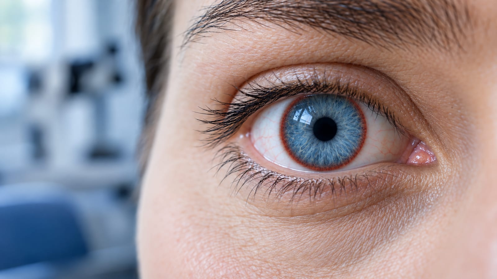
What Causes a Red Ring Around the Eye Pupil?
Not every red ring signals the same problem. Here are the most common causes, from urgent to benign:
Iritis (anterior uveitis). Inflammation of the iris produces a characteristic redness concentrated around the border of the iris and cornea, called a circumcorneal flush or peripupillary ring. This is the most medically significant cause and the focus of this article.
Conjunctivitis (pink eye). Redness spreads across the white of the eye rather than forming a tight ring around the iris. Usually accompanied by discharge and itching, rarely painful in the deep sense iritis is.
Corneal irritation or edema. A scratched cornea or corneal edema can cause limbal redness that mimics the ring pattern of iritis. An eye exam can quickly distinguish the two.
Limbal ring (cosmetic). A dark or pigmented ring at the outer edge of the iris is a normal anatomical feature, not redness or inflammation. This is sometimes called a limbal ring and is associated with youth and iris contrast. It requires no treatment.
Central heterochromia. A color variation within the iris itself, not a red ring, though it can alarm people who notice it for the first time. Completely benign.
Contact lens overuse: Prolonged wear can cause limbal vessel inflammation that mimics iritis.
If your ring is red, inflamed, or accompanied by pain, light sensitivity, or blurred vision, iritis is the most likely culprit and warrants urgent care.
What Is Iritis?
The iris, the colored ring around your pupil, sits in the anterior chamber at the front of the uvea, the eye’s middle layer. When it becomes inflamed, the condition is called iritis, or anterior uveitis. Inflammation of the iris disrupts the delicate structures of the anterior chamber, raising intraocular pressure and allowing inflammatory proteins to cloud the eye’s fluid, a finding ophthalmologists describe as “flare and cells” on slit-lamp examination.
Iritis can be acute, resolving within weeks with proper treatment, or chronic, persisting for months or years. Recurrent iritis, episodes that clear and then return, is also common, particularly in patients with an underlying autoimmune condition.
Red Ring From Iritis vs. a Limbal Ring or Heterochromia
Because search results often surface questions about limbal rings alongside iritis, it is worth clarifying the difference. A limbal ring is the naturally dark circle that outlines the iris where it meets the white of the eye. It is a cosmetic feature, not a medical symptom, and it requires no treatment. Central heterochromia, where the inner ring of the iris is a different color than the outer ring, is similarly a benign pigmentation variation. Neither of these involves redness, pain, or inflammation. Iritis, by contrast, produces a visible red or pink ring of dilated blood vessels encircling the cornea, a pattern called ciliary flush, and it is almost always accompanied by at least one of the following: aching eye pain, sensitivity to light, a constricted or irregularly shaped pupil, or blurred vision. If your ring is red rather than dark or multicolored, and if it appeared suddenly, that distinction matters enormously and warrants a same-day or next-day evaluation by an ophthalmologist.
What are the Symptoms of Iritis?
If you have noticed a red ring around your eye’s iris or pupil, you are likely looking at what doctors call a circumcorneal flush – a visible ring of inflamed, congested blood vessels at the limbus, the border where your cornea meets the white of your eye. This is one of the most recognizable and telling signs of iritis, and it often brings patients in for an urgent evaluation. Unlike general redness that spreads across the white of the eye, this ring-shaped flush sits specifically around the iris and pupil, making it visually distinct and clinically significant. If you see this in the mirror, your eye is signaling that something deeper than surface irritation is going on.
Iritis typically affects one eye at a time and comes on quickly. Additional symptoms include:
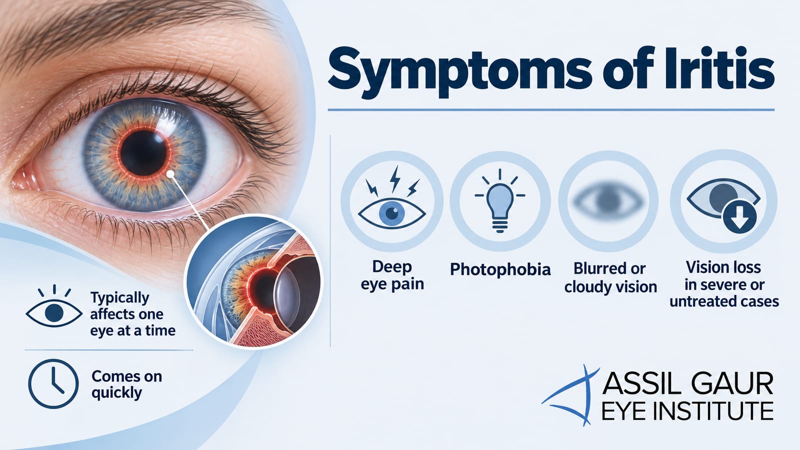
Deep eye pain (distinct from the surface irritation of conjunctivitis)
Photophobia (significant light sensitivity)
Blurred or cloudy vision
Vision loss in severe or untreated cases
In advanced cases, a visible white or yellowish layer of inflammatory cells at the bottom of the anterior chamber, called a hypopyon
A red ring around the iris is not always a sign of disease. A limbal ring is a naturally occurring dark circle at the outer edge of the iris and is simply a cosmetic feature of the eye’s anatomy; it requires no treatment and causes no discomfort. Similarly, central heterochromia, which creates a ring of a different color around the pupil, is a harmless variation in iris pigmentation. What sets iritis apart is the combination of redness, pain, and light sensitivity. If your red ring is accompanied by any discomfort or vision changes, that is a clinical symptom, not a cosmetic trait, and it warrants prompt evaluation by an ophthalmologist.
Does Iritis Have Other Complications?
Your eyes are among your most delicate and important structures. If iritis goes untreated, ongoing inflammation in the anterior chamber can cause damage that extends well beyond the iris. Untreated iritis can progress to serious complications, including:
Synechiae (adhesions between the iris and lens)
Macular edema (swelling at the back of the eye)
Elevated intraocular pressure and glaucoma
Retinitis (inflammation of the retina)
Early evaluation is the single most effective way to prevent these outcomes. If you are experiencing any of the symptoms above, contact us immediately rather than waiting to see if they resolve on their own.
What Are the Causes of Iritis?
Many cases of iritis are idiopathic, meaning no specific cause is identified. When a cause is found, it typically falls into one of these categories:
Eye trauma and injury. A blow to the eye, a penetrating injury, or even surgical trauma can trigger inflammation of the iris. This happens because physical damage disrupts the blood-ocular barrier, prompting an immune response inside the eye. The red ring around the eye pupil that follows trauma can appear within hours and should always be evaluated promptly. Traumatic iritis is one of the more common acute presentations seen in urgent eye care settings.
Infections. Herpes viruses (cold sores, shingles), Lyme disease, tuberculosis, toxoplasmosis, and syphilis are all linked to uveitis.
Genetics and autoimmune conditions. Patients carrying the HLA-B27 gene variant are significantly more likely to develop iritis, often with systemic autoimmune diseases such as ankylosing spondylitis, reactive arthritis, inflammatory bowel disease, and psoriatic arthritis.
Behçet’s disease. An uncommon cause in Western countries, also characterized by joint problems and oral or genital sores.
Juvenile idiopathic arthritis. Chronic iritis can develop silently in children with this condition.
Sarcoidosis. This autoimmune disease causes clusters of inflammatory cells throughout the body, including the eyes.
Medication-induced uveitis. Certain systemic medications, including some antibiotics and bisphosphonates, are associated with drug-induced iritis as a rare side effect. If you recently started a new medication and notice a red ring around your pupil alongside light sensitivity or blurred vision, see your eye doctor right away, as the timeline can be an important diagnostic clue.
Because so many different triggers can produce the same telltale redness and discomfort, pinpointing the underlying cause requires a thorough clinical evaluation rather than guesswork. The experienced ophthalmologists at Assil Gaur Eye Institute use advanced diagnostic tools to identify what is driving your inflammation and build a treatment plan around the root cause, not just the symptoms.
How Is Iritis Diagnosed?
Our ophthalmologists diagnose iritis through a thorough eye examination and medical history review. A slit-lamp examination is the cornerstone of diagnosis: this specialized microscope lets our doctors visualize the anterior chamber in detail, identify flare and cells in the aqueous humor, evaluate iris inflammation, and check for corneal edema or abrasions. Doctors often add dilating eye drops to examine the posterior segment. Doctors may order blood tests or imaging to identify an underlying autoimmune condition or infection.
Call now or book your appointment online.
Conjunctivitis vs. Iritis
Both pink eye (conjunctivitis) and iritis cause a red eye, but they behave very differently and require very different responses. If you notice a red ring around your pupil or a circumcorneal flush concentrated near the iris, that pattern points strongly toward iritis rather than conjunctivitis. Here is how to read the signs.
Onset and discharge. Conjunctivitis tends to arrive quickly, often overnight, and almost always produces discharge, either watery and clear or thick and purulent. You may wake up with your eyelids stuck together. Iritis, by contrast, builds more gradually over hours to a day or two, and produces no discharge at all. A red, irritated eye with no discharge and no crusting is a meaningful clue that something deeper than the surface is inflamed.
Pain and light sensitivity. Conjunctivitis causes surface irritation, a gritty or scratchy feeling, but not deep pain. Iritis causes dull, aching pain behind the eye and almost always comes with significant photophobia. Bright light can feel genuinely painful, not just uncomfortable. If stepping into sunlight or a lit room makes you wince and reach for sunglasses, that level of light sensitivity is a red flag that warrants same-day evaluation.
Vision and contagion. Conjunctivitis rarely affects how clearly you see, and it is highly contagious. Iritis is not contagious, but it can blur or reduce vision as inflammation builds inside the anterior chamber. Treat any combination of deep eye pain, light sensitivity, blurred vision, and a red ring concentrated around the limbus rather than spread across the white of the eye as urgent. Do not wait to see whether it resolves on its own, because untreated iritis can lead to serious complications including synechiae, elevated eye pressure, and permanent vision loss.
When in doubt, pain and photophobia strongly favor iritis over conjunctivitis, and a same-day ophthalmology appointment is the right call. The world-class team at Assil Gaur Eye Institute is equipped to evaluate both conditions with precision and get you the answers you need quickly.
Iritis or Uveitis?
Iritis is a form of uveitis because the iris is part of the uvea, the eye’s middle vascular layer. The uvea includes the iris, the ciliary body, and the choroid. Inflammation in each zone has its own name:
Anterior uveitis: Iritis, the most common form, affecting the iris at the front of the uvea.
Intermediate uveitis: Inflammation of the vitreous gel in the middle of the uvea.
Posterior uveitis: Also called chorioretinitis, involving the choroid at the back of the uvea.
The circumcorneal flush, the visible red ring around the iris, is a hallmark sign of anterior uveitis and one of the clearest indicators that the inflammation originates inside the eye rather than on its surface. When patients search for answers about a red ring around the eye or pupil, anterior uveitis most consistently matches that description, which is why prompt evaluation by an ophthalmologist matters. Unlike surface-level redness from allergies or a broken blood vessel, a circumcorneal flush signals deeper involvement and requires prompt treatment to protect your vision.
Iritis Treatment
Treatment is effective when started promptly. The standard approach includes:
Steroid eye drops. The first-line treatment for iritis almost always involves two types of prescription eye drops used together. Corticosteroid eye drops, typically prednisolone acetate, are prescribed to calm the inflammation directly at its source. These are usually applied frequently at first, sometimes every one to two hours during the acute phase, and then tapered gradually over several weeks as the eye responds. Stopping too soon is one of the most common reasons iritis flares back up, so following your ophthalmologist’s tapering schedule matters.
Cycloplegic or mydriatic drops. The second type of drop is a cycloplegic agent, such as atropine or cyclopentolate. These dilate the pupil and temporarily paralyze the ciliary muscle, which does two important things: it relieves the deep, aching pain caused by ciliary spasm, and it prevents the iris from sticking to the lens of the eye. If they form, those adhesions, called posterior synechiae, can distort the pupil and dangerously raise eye pressure. Cycloplegic drops are a critical part of treatment that is sometimes overlooked in general discussions of iritis care.
Monitoring intraocular pressure. Steroid drops can raise eye pressure, so follow-up monitoring appointments are essential throughout treatment.
Oral steroids or immunosuppressants. Severe or recurrent iritis, particularly in patients with an identified autoimmune condition, may require systemic treatment to control the underlying inflammation. This can include oral corticosteroids and immunosuppressants such as methotrexate or mycophenolate. Rheumatologists typically manage these systemic medications, especially when iritis is linked to conditions like ankylosing spondylitis or HLA-B27 positivity. Treating the root cause is as important as treating the eye itself, because recurrent iritis without an identified trigger can cause cumulative damage over time.
Treating the root cause. When iritis is linked to an infection or systemic disease, addressing that condition directly is essential to preventing recurrence.
Most acute cases resolve within six to eight weeks with appropriate treatment. Chronic or recurrent iritis requires longer-term management and closer monitoring by a specialist.
Why Choose Assil Gaur Eye Institute for Iritis Care in Los Angeles?
At Assil Gaur Eye Institute, our world-class ophthalmologists bring advanced diagnostic precision and personalized care to every patient who walks through our doors, including those dealing with iritis and other inflammatory eye conditions. From slit-lamp evaluation to identifying underlying systemic causes, our team takes a thorough, expert approach that goes far beyond a surface-level exam. Patients across Los Angeles and Southern California trust us for the best eye care, and we are here to guide you from your first symptom to full recovery.
Schedule your complimentary evaluation by calling 866-945-2745 or requesting an appointment online. Our clinics serve patients throughout Beverly Hills, Santa Monica, West Los Angeles, West Hollywood, Culver City, Hollywood, Venice, Marina del Rey, Malibu, Manhattan Beach, and Downtown Los Angeles. Every eye is precious at Assil Gaur Eye Institute, and yours deserves world-class attention.
Call now or book your appointment online.

Written by The AGEI Educational Team

Medically Reviewed by Dr. Kerry Assil