What is a conjunctival nevus?
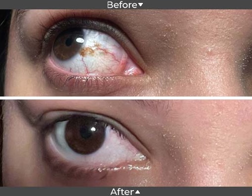
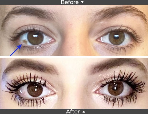
A conjunctival nevus is a freckle or mole-like spot that often appears on the transparent film covering the eye. Nevi (plural for nevus) can range from dark brown to yellow; over time, these nevi can darken or lighten in color. They are often located adjacent to the iris (the colored part of the eye).
Nevi are made up of pigment cells, called melanocytes, that provide color to your skin, hair, and eyes. When several of the melanin cells cluster together, they form a nevus.
They are commonly found in Caucasians because light eyes, fair skin, and those who burn easily when sun exposure are also risk factors.
- What causes eye freckles?
- What are the symptoms of conjunctival nevus?
- How worried should I be about an eye freckle?
- When should you see your eye doctor about an eye freckle (nevus)?
- What are treatments for conjunctival nevus?
- Eye freckle removal surgery
- Why choose Assil Gaur Eye Institute for your eye freckle removal
- Eye Freckle FAQs
- Are there different types of nevi?
What causes eye freckles?
Large-scale studies have found a link between chronic sun exposure and the incidence of nevi. The American Academy of Ophthalmology (AAO) suggests that exposure to ultraviolet (UV) light may contribute to the formation of choroidal nevi.
A family history of nevi does not play a role in determining your likelihood of developing conjunctival pigmentation.
What are the symptoms of conjunctival nevus?
As with freckles anywhere on your body, conjunctival nevi aren't associated with pain or discomfort. Although nevi tend to remain stable, they can grow due to inflammation or hormonal changes.
Honestly, the most common symptom of an eye freckle for our patients is emotional discomfort or insecurity. Since people often look us in the eye when we speak with them, it is much more noticeable than a freckle on your hand or a mole on the back of your neck.
How worried should I be about an eye freckle?

A nevus is rarely cancerous (conjunctival melanoma), and people are often born with harmless eye nevi (benign melanocytic tumor or lesion). A nevus that develops later in life is typically also harmless, but, like a skin mole, it could develop into eye cancer (called ocular melanoma). Again, though rare, it should be monitored with periodic eye exams.
When should you see your eye doctor about an eye freckle (nevus)?
If you notice something that looks like a freckle in your eye that wasn’t there before, you should get it checked by an ophthalmologist. Although it’s likely harmless, your eye care provider will want to examine it closely and continue regular checkups to ensure it doesn’t change.
There could be other issues if you have a nevi and are experiencing:
- Blurry vision or other vision changes.
- Eye floaters or flashing lights
- Eye pain or discomfort
- Changes in the size or color of the freckle.
These can be symptoms of retinal detachment. Choroidal nevi can sometimes leak fluid, resulting in abnormal blood vessels, which can lead to retinal detachment and vision loss.
What are treatments for conjunctival nevus?
Because nevi are painless and don't affect vision, they don’t necessarily require treatment other than periodic observation to monitor for changes in the nevus over time.
Our doctors will thoroughly examine your eye's front and inside structures, including the retina, the macula, and the optic nerve.
Just as with skin cancer, during your eye examination, your doctor will look for the ABCDE clinical features of moles that warrant close monitoring:
- Asymmetrical shape
- Irregular borders
- Color changes or several colors present
- Diameter that has advanced
- Evolution of the nevi's appearance over time
During your examination, our doctors will direct our highly trained staff to take pictures of the nevus and compare them over time to see if it has changed in size or shape. You may be asked to return to have the nevus re-checked in six months.
If the nevus does not change over a year or two, it is unlikely to be choroidal melanoma. However, our ophthalmologists recommend checking it regularly since it is possible for it to become something serious.
Schedule your consultation today with the internationally recognized conjunctival nevus specialists at AGEI
Eye freckle removal surgery

Assil Gaur Eye Institute removes conjunctival nevi for two common reasons:
- If melanoma is suspected: If there are any cancer concerns, our eye doctor will recommend an excisional biopsy, where the pigmented lesion is removed surgically. The removed tissue is sent to a pathology lab to assess the presence of any cancer cells.
- Cosmetic reasons: Thanks to techniques developed by renowned ophthalmologist Dr. Kerry Assil, removing a cosmetic nevus is now a much simpler and gentler procedure than it used to be.
The procedure involves using a very mild thermal brushing technique to extract the pigment from the surface of the conjunctiva. Recovery is typically quick and painless, with no trace of the prior nevus. Unfortunately, because this treatment is considered cosmetic, insurance does not typically cover it.
Why choose Assil Gaur Eye Institute for your eye freckle removal
The team of ophthalmologists at AGEI have performed thousands of eye freckle removal surgeries and are nationally recognized leaders in the field, thanks to their proprietary removal techniques.
Our nationally recognized ophthalmologists and eye institutes are leaders in a wide range of ophthalmological conditions, including state-of-the-art LASIK vision correction, retinal treatments, cataract surgery, glaucoma care, macular disease, and, of course, diabetic eye conditions.
This is one reason why Los Angeles Magazine named Assil Gaur Eye Institute one of the Top ophthalmology doctors in Los Angeles year after year.
At AGEI, you will experience a state-of-the-art healthcare facility that combines revolutionary technologies with experienced vision care professionals. Our goal is to help you achieve your best vision and optimal eye health.
We are conveniently located for patients throughout Southern California and the Los Angeles area in or near Beverly Hills, Santa Monica, West Los Angeles, West Hollywood, Culver City, Hollywood, Venice, Marina del Rey, Malibu, Manhattan Beach, and Downtown Los Angeles.
Eye Freckle FAQs
Are there different types of nevi?
There are different types of nevi based on where the nevus is located.
Conjunctival nevus
This type of eye freckle is the conjunctiva, the transparent film covering the front of your eyes. It usually shows up because it appears on the sclera or the white part of your eye. Conjunctival nevi are the common type of nevi.
Iris nevus and iris freckle
An iris nevus and iris freckles appear in the iris. An iris freckle is smaller than an iris nevus. However, an iris nevus goes deeper into the iris and sometimes can pull the pupil to the side.
Choroidal nevus
A choroidal nevus occurs in the choroid, the layer of tissue beneath the retina in the back of the eye. The choroid is part of the uvea, the pigmented part of your eye, and includes the iris. Both choroidal nevi and iris nevi are forms of uveal nevi.
Resources
Conjunctival Nevi: Clinical Features and Natural Course in 410 Consecutive Patients
https://doi.org/10.1001/ARCHOPHT.122.2.167
Surgical Outcome of Chemical Peeling of Conjunctival Nevus with Alcohol
https://doi.org/10.3341/jkos.2016.57.5.705
Conjunctival naevi in Denmark 1960-1980. A 21-year follow-up study
https://www.ncbi.nlm.nih.gov/pubmed/8883545
Conjunctival lesions in adults. A clinical and histopathologic review
https://www.ncbi.nlm.nih.gov/pubmed/3301209
Subepithelial conjunctival nevus with atypia: Expanding our understanding of a challenging diagnosis
https://www.sciencedirect.com/science/article/pii/S2214330018300300
-
Kerry K. Assil, MD, is regarded as one of the world’s foremost experts in refractive surgery, having made significant advances in the field with his numerous inventions. Additionally he has the unique distinction of having trained thousands of eye surgeons in the latest refractive surgical techniques.
Dr. Assil has authored more than one hundred textbooks, textbook chapters and articles on refractive surgery and has appeared regularly on major television network news programs as a pioneer in refractive surgery. He also leads educational forums for other eye care professionals, which have included featured lectureships at Harvard University, Johns Hopkins University and Tokyo University.


