 Wh
WhWhat is keratoconus?
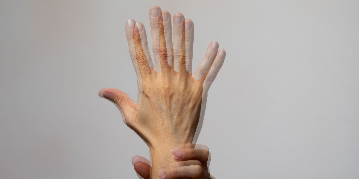
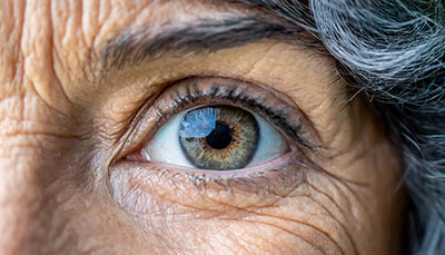
Keratoconus is an eye condition in which the cornea (the clear outer layer of the eye) progressively thins and becomes irregularly (coned) shaped.
The abnormal curvature of the cornea causes the cornea to bulge and prevents the light entering the eye from focusing correctly on the retina, and causes vision to become increasingly distorted. As the condition worsens and the front of the cornea expands, you begin to experience myopia(nearsightedness) and develop irregular astigmatism.
- What are the symptoms of keratoconus?
- What causes keratoconus?
- Keratoconus risk factors
- How is keratoconus diagnosed?
- What are treatments for early keratoconus?
- What are the treatments for late-stage keratoconus?
- Do all keratoconus patients eventually need a cornea transplant?
- Why is Assil Gaur Eye Institute the best keratoconus specialist near me?
- Keratoconus FAQs
- How common is keratoconus?
- Can LASIK help keratoconus?
- How do you find the best keratoconus specialist near me?
What are the symptoms of keratoconus?
Signs and symptoms of keratoconus can include:
-
Blurred vision or distorted vision with difficulty seeing near and far
-
Increasing light sensitivity and glare, and halos, which can compromise night driving
-
Unstable vision leading to frequently changing eye prescriptions
-
Increased dry eye symptoms while wearing conventional contact lenses.
What causes keratoconus?
Keratoconus is more prevalent in teenagers and adults in their 30s. It can progress gradually or quickly and usually affects both eyes. Unfortunately, the cause of keratoconus is not fully understood. There is a known genetic connection since ten percent of keratoconus sufferers have a parent with the condition. It’s also often seen in patients who suffer from retinitis pigmentosa, Down syndrome, Ehlers-Danlos syndrome, hay fever, and asthma.
Some medical researchers theorize that an enzyme imbalance within the cornea makes it vulnerable to damage from free radicals, which causes the cornea tissue to weaken and bulge.
Keratoconus risk factors
Frequent vigorous eye rubbing is thought to increase your risk of developing keratoconus. Additionally, overexposure to ultraviolet rays, improper contact lens wear, and chronic eye irritations may increase your odds of developing keratoconus.
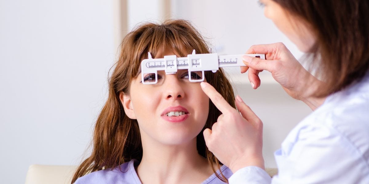
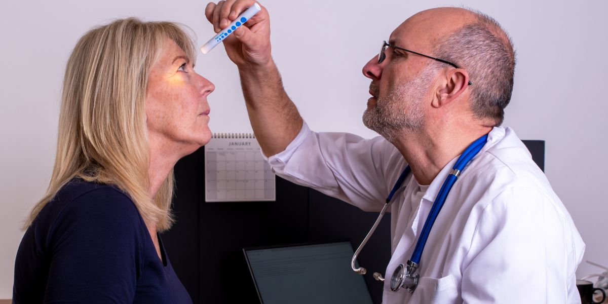
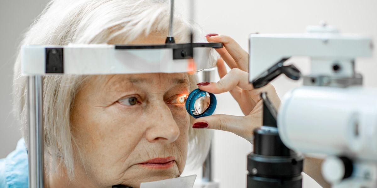
How is keratoconus diagnosed?
Typically, keratoconus is diagnosed during an eye exam when our doctors observe the irregularly shaped cornea.
As part of a comprehensive eye exam, our eye doctor measures the shape of your cornea to screen for keratoconus. By performing annual eye exams, we can detect and track subtle changes in the early keratoconus stages.
At AGEI, we use state-of-the-art corneal topography, which involves imagining the cornea to analyze its surface shape and characteristics in just a few seconds.
Schedule your consultation today with our internationally recognized doctors
What are treatments for early keratoconus?
There are several stages of keratoconus, starting with early diagnosis. The earlier you are diagnosed, the greater the chance we can often easily correct visual issues with glasses or special contact lenses.
Early treatment options, which can often provide excellent quality vision and avoid vision loss for many years, include:
Rigid gas-permeable keratoconus lenses
Rigid gas-permeable lenses are made of specialized plastic that transmits oxygen. These lenses reshape your cornea’s curvature to correct your vision while wearing them.
Custom Toric soft contact lenses
While these are usually more comfortable than rigid gas-permeable contact lenses, they are best for individuals with more astigmatism.
Scleral contact lenses
These are oversized contact lenses whose outer rim extends to the white of your eye, known as the sclera. These lenses vault over the cone-shaped part of your cornea without touching its surface due to a special fluid-filled reservoir that lies over the cone-shaped cornea, restoring clear vision comfortably.
Finding properly fitting corrective lenses is crucial. Poorly fit lenses can result in corneal scarring and intolerance of any lens.
At AGEI, we have experts who can help identify the lens option that fits your needs and provide you with the best vision and comfort.
Corneal Collagen Crosslinking for keratoconus
The progression of keratoconus can be treated with early detection and a treatment called Collagen Crosslinking (CXL). This FDA-approved technology is the only kind designed to reduce the progression of keratoconus.
Cross-linking collagen involves activating riboflavin (vitamin B) using ultraviolet light to generate collagen bonds between corneal fibers. This treatment can strengthen your cornea by up to 300%. By strengthening the cornea, the bulging process is slowed down.
Studies have found that combining keratoconus contact lens wear with corneal cross-linking appears to be even more effective in avoiding transplantation surgery than either treatment alone.
Intacs
Intacs is a keratoconus treatment introduced over 20 years ago that involves placing concentric plastic rings under the corneal surface to flatten it and improve vision.
While AGEI was one of the principal investigators during the initial FDA trials for Intacs, we’ve since stopped offering this procedure to our patients.
What are the treatments for late-stage keratoconus?
In up to twenty percent of keratoconus patients, the cornea eventually becomes so scarred that they can no longer tolerate wearing contact lenses. At this point, a corneal transplant may be the only way to preserve vision.
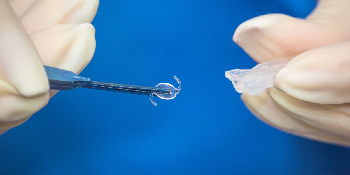
Corneal Transplantation
Corneal transplantation (or Penetrating Keratoplasty) is an outpatient eye surgery that offers Keratoconus patients excellent results.
The procedure involves removing the abnormal corneal tissue from the eye and replacing it with a graft of healthy corneal tissue from a cadaver donor. The donor cornea graft is held in place with stitches one-third the width of a human hair.
Our patients usually get excellent results with a cornea transplant, although their vision may stay blurry for several months following surgery. To ensure the best possible outcome, you will be placed on medication, including eye drops, to prevent graft rejection following a transplant. In most cases, you will likely continue to need eyeglasses or contact lenses to achieve your best vision.
Learn more about cornea transplants.
Do all keratoconus patients eventually need a cornea transplant?
No! Early detection and treatments like corneal cross-linking and therapeutic contact lenses can often successfully treat keratoconus and restore visual acuity. These early treatments have proven to be very effective in slowing or stopping the progression of keratoconus.
Why is Assil Gaur Eye Institute the best keratoconus specialist near me?
The Assil Gaur Eye Institute team of keratoconus doctors is Southern California’s Leader in Keratoconus Treatment. They have extensive experience in keratoconus corneal eye disease and its management. Dr. Assil's leadership in corneal transplantation surgery began while serving on the university faculty in St. Louis decades ago in one of the largest corneal transplant practices in the USA.
Dr. Assil and his team have performed thousands of corneal transplants and are nationally recognized leaders in the field.
The doctors at AEGI ophthalmology group have a combined 100 years of healthcare experience and are nationally recognized specialists in treating glaucoma, macular degeneration, and diabetic retinopathy.
This is one of the reasons that Los Angeles Magazine named Assil Gaur Eye Institute as one of the Top ophthalmology eye centers in Los Angeles, year after year. Optometrists from all over Southern California refer their patients to us for care.
We are conveniently located for patients throughout Southern California and the Los Angeles area in or near Beverly Hills, Santa Monica, West Los Angeles, West Hollywood, Culver City, Hollywood, Venice, Marina del Rey, Malibu, Manhattan Beach, and Downtown Los Angeles.
![]()
Keratoconus FAQs
How common is keratoconus?
While keratoconus is the most common cornea ectatic disorder, it affects about 5% of the population worldwide.
Can LASIK help keratoconus?
No, LASIK is not recommended for keratoconus patients. The reasons include:
- The cornea in keratoconus is already weak and unstable, and LASIK can further compromise its structural integrity.
- The irregular shape of the cornea in keratoconus makes accurate measurement and correction challenging, leading to unpredictable and potentially unsatisfactory outcomes.
- LASIK does not address the underlying progression of keratoconus, which may render the corrective procedure ineffective as the condition advances.
How do you find the best keratoconus specialist near me?
- Seek Referrals: Start by asking for recommendations from your primary eye care provider or ophthalmologist. They can provide valuable insights and refer you to specialists experienced in managing keratoconus.
- Research Credentials: Look for ophthalmologists or cornea specialists with specific expertise in diagnosing and treating keratoconus. Check their credentials, education, and certifications. Board certification in ophthalmology and memberships in professional organizations can be good indicators of their expertise.
- Experience and Specialization: Consider doctors with significant experience in managing keratoconus and related corneal conditions. Look for specialists who focus on the cornea and external disease or refractive surgery.
Sources:
Chou, B., Onofrey B. "Keratoconus." In Ocular Therapeutics Handbook: A Clinical Manual (Fourth Edition), edited by Onofrey, B, Skorin L, Holdeman N, 700-704...
Keratoconus articles from our blog
-

Kerry K. Assil, MD, is regarded as one of the world’s foremost experts in refractive surgery, having made significant advances in the field with his numerous inventions. Additionally he has the unique distinction of having trained thousands of eye surgeons in the latest refractive surgical techniques.
Dr. Assil has authored more than one hundred textbooks, textbook chapters and articles on refractive surgery and has appeared regularly on major television network news programs as a pioneer in refractive surgery. He also leads educational forums for other eye care professionals, which have included featured lectureships at Harvard University, Johns Hopkins University and Tokyo University.

 Dr. Adele was outstanding. My eye was in bad shape and as soon as she started taking a look and talking me through what was goi...Michael P.
 Their whole staff is fabulous and so caring!!! The Annual Eye Exam that Dr May Performs is so extensive and done with such...Joan M.
- I am pleased with the service I have received. And I will continue to recommend to my friends and familyFelix C.
- The staff is personable and professional.Kurt E.
- Professional courteous. I am thankful for the care I was given. I would recommend Dr Adeleh to friends who are seeking competen...Michael G.
- Dr may is greatJennifer L.
- Very good treatment!Steven M.
- NopeCaroline M.
- Always too long of a waitCindra L.
- MD was very attentive to my concerns.Charles D.
- You can't possibly make a better choice than Dr assil and his staff. They are the absolute best.Natural R.
- Everything was great, not much else I can sayBarbara M.
-
 Listen Now
Are Cataracts at a Young Age Getting More Common?
Read
Listen Now
Are Cataracts at a Young Age Getting More Common?
Read
-
 Listen Now
What Are the Causes of Double Vision and How Can They Be Addressed?
Read
Listen Now
What Are the Causes of Double Vision and How Can They Be Addressed?
Read
-
 Listen Now
The History of IOLs: You’ve Come A Long Way, Baby!
Read
Listen Now
The History of IOLs: You’ve Come A Long Way, Baby!
Read
-
 Listen Now
Cataract Lens Replacement Failure: What Can You Do About It?
Read
Listen Now
Cataract Lens Replacement Failure: What Can You Do About It?
Read
-
 Listen Now
Can Pupil Size Reveal Intelligence? Maybe Size Really Does Matter!
Read
Listen Now
Can Pupil Size Reveal Intelligence? Maybe Size Really Does Matter!
Read
-
 Listen Now
Understanding Pinguecula: Causes, Symptoms, and Treatment
Read
Listen Now
Understanding Pinguecula: Causes, Symptoms, and Treatment
Read
-
 Listen Now
Alcohol and Dry Eye Syndrome: Understanding the Connection
Read
Listen Now
Alcohol and Dry Eye Syndrome: Understanding the Connection
Read
-
 Listen Now
What are Some Common Medications that can Cause or Worsen Glaucoma?
Read
Listen Now
What are Some Common Medications that can Cause or Worsen Glaucoma?
Read
-
 Listen Now
Can Vitamin D Help Relieve Dry Eye Disease? A New Study Says Yes!
Read
Listen Now
Can Vitamin D Help Relieve Dry Eye Disease? A New Study Says Yes!
Read
-
 How Autoimmune Diseases Affect Your Eyes: What You Need to Know
Read
How Autoimmune Diseases Affect Your Eyes: What You Need to Know
Read
-
 Breakthrough Eye Implant Technology Offers New Hope for Patients with Advanced Macular Degeneration
Read
Breakthrough Eye Implant Technology Offers New Hope for Patients with Advanced Macular Degeneration
Read
-
 Kerry and Tanaz Assil focus on the fruits of their labor
Read
Kerry and Tanaz Assil focus on the fruits of their labor
Read
-
 Listen Now
Patients are raving about the enVista® Envy™ Trifocal IOL Lens
Read
Listen Now
Patients are raving about the enVista® Envy™ Trifocal IOL Lens
Read
-
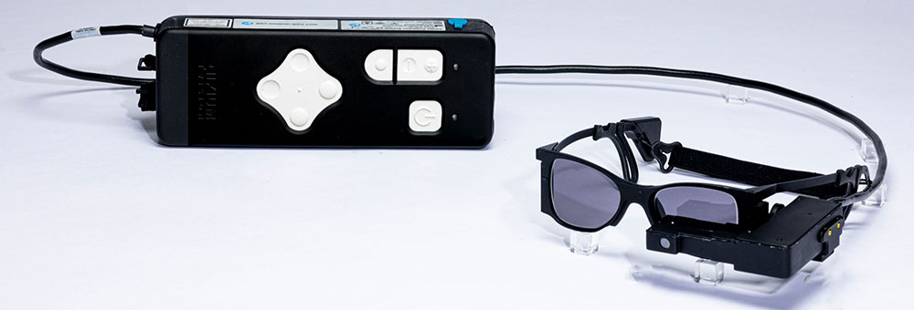
 Valeda Light Delivery System for Dry Age-Related Macular Degeneration (AMD)
Read
Valeda Light Delivery System for Dry Age-Related Macular Degeneration (AMD)
Read
-
 Listen Now
Can Stress Increase Eye Pressure in Glaucoma Patients?
Read
Listen Now
Can Stress Increase Eye Pressure in Glaucoma Patients?
Read


Look Who Trusts Their Eyes to Assil Gaur Eye Institute...
-
LeBron James
Los Angeles LakersLeBron James is one of the NBA's greatest of all time. He absolutely depends on his vision to perform at the very highest level. That's why he's an AGEI LASIK surgery patient.
-
Anthony Davis
Dallas Mavericks -
Chris Paul
San Antonio Spurs -
Paul George
Philadelphia 76ers -
Marlee Matlin
Actress -
Michelle Williams
Actress -
Dwyane Wade
Miami Heat (retired) -
LaToya Jackson
Entertainer -
Lorenzo Lamas
Actor -
Philip Bailey
Earth Wind and Fire -
Gary Sinise
Actor -
Rip Hamilton
Former NBA player -
John Salley
Commentator, former NBA player -
Blair Underwood
Actor -
Tom ArnoldComedian/Actor
-
Troy EvansActor
-
John NobleActor
-
Isaac EddyActor/Director/Teacher
-
Barbara RushActress
-
John EmersonUS Ambassador to Germany
-
Shane MosleyFormer professional boxer
-
Cuttino MobleyFormer NBA player
-
Seth GordonDirector
-
Maurice EvansFormer NBA player
-
Wang LuoyongActor
-
Jesse MetcalfeActor
-
Ananda LewisTelevision Personality
-
Rob BrydonComedian
-
Clifton Collins Jr.Actor
-
Malinda WilliamsActress
-
Adrienne FrantzActress
-
Brad PittActor
-
LeBron JamesLos Angeles Lakers
-
Anthony DavisDallas Mavericks
-
Chris PaulSan Antonio Spurs
-
Courtney CoxActress
-
Paul GeorgePhiladelphia 76ers
-
Marlee MatlinActress
-
Michelle WilliamsActress
-
Dwyane WadeMiami Heat (retired)
-
Jalen BrunsonNew York Knicks
-
Mychal ThompsonCommentator, former NBA player
-
LaToya JacksonEntertainer
-
Lorenzo LamasActor
-
Philip BaileyEarth Wind and Fire
-
Gary SiniseActor
-
Rip HamiltonFormer NBA player
-
John SalleyCommentator, former NBA player
-
Blair UnderwoodActor
-
Tom ArnoldComedian/Actor
-
Troy EvansActor
-
John NobleActor
-
Isaac EddyActor/Director/Teacher
-
Barbara RushActress
-
Rudy GarciduenasBlue Man Group
-
John EmersonUS Ambassador to Germany
-
Shane MosleyFormer professional boxer
-
Cuttino MobleyFormer NBA player
-
Seth GordonDirector
-
Maurice EvansFormer NBA player
-
Cheri BlauwetPhysician and Wheelchair Racer
-
Wang LuoyongActor
-
Jesse MetcalfeActor
-
Jeana WilsonActress
-
Courtnee DraperActress
-
Ananda LewisTelevision Personality
-
Robyn JohnsonProducer
-
Rob BrydonComedian
-
Cary SchumanActor
-
Roland Buddy Lewis Jr.Comedian
-
Clifton Collins Jr.Actor
-
Malinda WilliamsActress
-
Chad JeffersMusician
-
Adrienne FrantzActress
-
Franco CarlottoBody Builder
-
Brad MatesMusician
-
David PichetteMusician
-
Mike MelaconProfessional Baseball Player
-
Nautica de la CruzRadio Personality
-
Lynn ConkwrightBody Builder
-
Nely GalanProducer
-
Roxanne GallaActress